nid: 63813
Additional formats:
None available
Description:
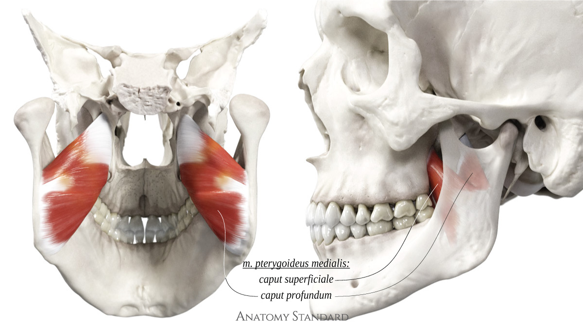
Medial pterygoid muscles: posterior and lateral view. The medial pterygoid muscle has two heads: the superficial head is smaller and arises from the maxilla, while the deeper head is larger and originates from the pterygoid fossa. Origin: fossa pterygoidea (medial head) + tuber maxillae (lateral head). Insertion: tuberositas pterygoidea maxillae. Function: elevatation of the mandible. It also contributes to limited protrusion. When acting unilaterally, it assists in moving the mandible to the contralateral side. Latin labels.
Image (CC BY-NC) and description retrieved from Anatomy Standard, page Masticatory Muscles.
Image (CC BY-NC) and description retrieved from Anatomy Standard, page Masticatory Muscles.
Anatomical structures in item:
Uploaded by: rva
Netherlands, Leiden – Leiden University Medical Center, Leiden University
Musculus pterygoideus medialis
Creator(s)/credit: Jānis Šavlovskis MD, PhD, Assistant Professor; Kristaps Raits, 3D generalist
Requirements for usage
You are free to use this item if you follow the requirements of the license:  View license
 View license
 If you use this item you should credit it as follows:
- For usage in print - copy and paste the line below:
- For digital usage (e.g. in PowerPoint, Impress, Word, Writer) - copy and paste the line below (optionally add the license icon):
"Anatomy Standard - Medial pterygoid muscle: posterior and lateral view - Latin labels" at AnatomyTOOL.org by Jānis Šavlovskis and Kristaps Raits, license: Creative Commons Attribution-NonCommercial


Comments