
nid: 63804
Additional formats:
None available
Description:
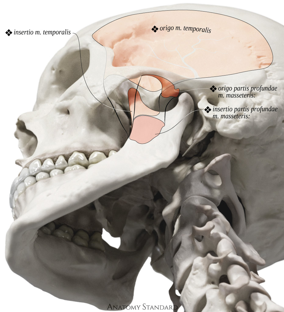
Temporal muscle and deep part of masseter muscle: inferolateral view. The masseter is the most prominent masticatory muscle. It consists of two clearly defined parts with differently directed fibers. The masseter muscle consists of the superficial part (Origin: zygomatic arch, mostly on the zygomatic bone; insertion: angle of mandible (masseteric tuberosity); function: elevate (close) lower jaw) and the deep part (Origin: zygomatic arch, mostly on the temporal bone & the capsule of the temporomandibular joint; insertion: ramus of mandible, mostly above the masseteric tuberosity; function: elevate (close) lower jaw, lateral stabilization of the discocapsular system). Latin labels.
Image and description retrieved from Anatomy Standard, page Masticatory Muscles.
Image and description retrieved from Anatomy Standard, page Masticatory Muscles.
Anatomical structures in item:
Uploaded by: rva
Netherlands, Leiden – Leiden University Medical Center, Leiden University
Musculus masseter
Pars profunda musculus masseterica
Musculus temporalis
Articulatio temporomandibularis
Creator(s)/credit: Jānis Šavlovskis MD, PhD, Assistant Professor; Kristaps Raits, 3D generalist
Requirements for usage
You are free to use this item if you follow the requirements of the license:  View license
View license If you use this item you should credit it as follows:
- For usage in print - copy and paste the line below:
- For digital usage (e.g. in PowerPoint, Impress, Word, Writer) - copy and paste the line below (optionally add the license icon):
"Anatomy Standard - Origo and insertio of temporal muscle and deep part of masseter muscle: anterolateral view - Latin labels" at AnatomyTOOL.org by Jānis Šavlovskis and Kristaps Raits, license: Creative Commons Attribution-NonCommercial


Comments