
nid: 63790
Additional formats:
None available
Description:
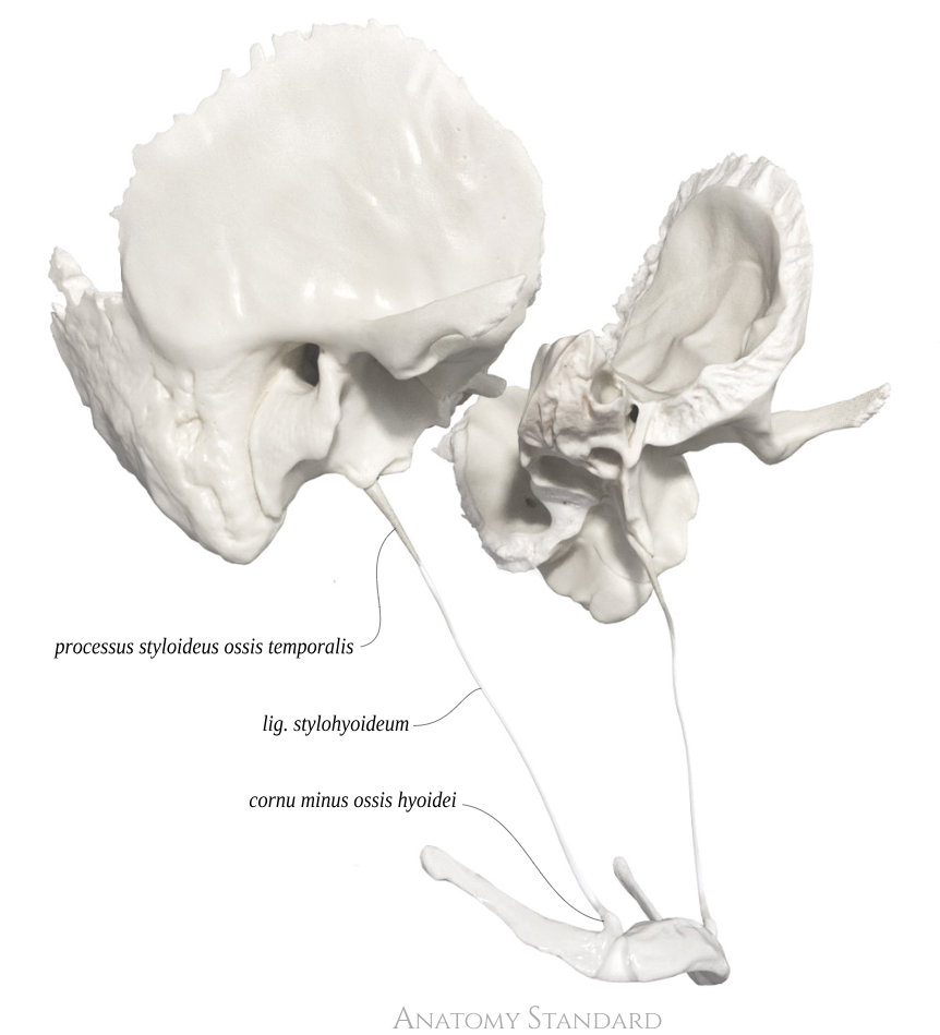
Ligamentum stylohyoideum: anterolateral view. The stylohyoid ligament connects the styloid process of the temporal bone with the lesser horn of the os hyoideum. All the aforementioned anatomical structures develop from the cartilage of the second pharyngeal (visceral) arch during embryogenesis. After birth, the cartilaginous structure transforms into the stylohyoid ligament, with ossification beginning at its ends, gradually elongating the styloid process on one side and forming part of the hyoid bone on the other. Latin labels.
Image and description retrieved from Anatomy Standard, page Cranial Syndesmoses.
Image and description retrieved from Anatomy Standard, page Cranial Syndesmoses.
Anatomical structures in item:
Uploaded by: rva
Netherlands, Leiden – Leiden University Medical Center, Leiden University
Ligamentum stylohyoideum
Processus styloideus
Cornu minus ossis hyoidei
Creator(s)/credit: Jānis Šavlovskis MD, PhD, Assistant Professor; Kristaps Raits, 3D generalist
Requirements for usage
You are free to use this item if you follow the requirements of the license:  View license
View license If you use this item you should credit it as follows:
- For usage in print - copy and paste the line below:
- For digital usage (e.g. in PowerPoint, Impress, Word, Writer) - copy and paste the line below (optionally add the license icon):
"Anatomy Standard - Ligamentum stylohyoideum: anterolateral view - Latin labels" at AnatomyTOOL.org by Jānis Šavlovskis and Kristaps Raits, license: Creative Commons Attribution-NonCommercial


Comments