nid: 63590
Additional formats:
None available
Description:
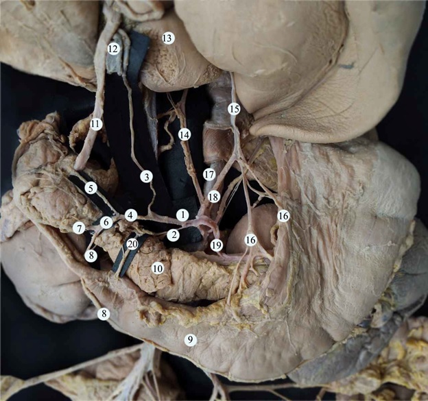
Vascular supply of liver, duodenum, pancreas, spleen, and stomach.
Abstract: we describe a case of unusual development of the celiac trunk observed in the cadaver of 1-year old male child. The celiac trunk branched into five vessels: the splenic, common hepatic and left gastric arteries, the left inferior diaphragmatic artery, and a short trunk that branched into the right inferior diaphragmatic artery and right accessory hepatic artery. Additionally, the manner of branching of the vessel was unusual: it was possible to distinguish two branching points that corresponded to its s-shaped trajectory. There were also other variations of vascular supply, such as the presence of a left accessory hepatic artery, an additional superior pancreatoduodenal artery, and others. It should be noted that multiple developmental variations can be common in clinical practice and clinicians should be aware of them during diagnostic and interventional procedures.
(1) common hepatic artery; (2) additional superior pancreatoduodenal artery; (3) proper hepatic artery; (4) gastroduodenal artery; (5) superior pancreatoduodenal artery; (6) common trunk; (7) the inferior artery of the pancreatic head; (8) right gastro-epiploic artery; (9) the stomach; (10) pancreas; (11) common bile duct; (12) portal vein; (13) the liver; (14) right accessory hepatic artery; (15) left accessory hepatic artery; (16) arteries to the lesser curvature of the stomach; (17) abdominal aorta; (18) left gastric artery; (19) splenic artery; (20) branch to the wall of the duodenal ampulla.
Image, abstract and description derived from Covantev S, Mazuruc N, Drangoi I, Belic O. Unusual development of the celiac trunk and its clinical significance J Vasc Bras. 20:2021;e20200032, licenced under Creative Commons Attribution (CC BY).
Anatomical structures in item:
Uploaded by: rva
Netherlands, Leiden – Leiden University Medical Center, Leiden University
Arteria hepatica communis
Arteria pancreaticoduodenalis superior anterior
Arteria hepatica propria
Arteria gastroduodenalis
Arteria pancreatica inferior
Arteria gastroomentalis dextra
Ventriculus
Pancreas
Ductus biliaris
Vena portae hepatis
Hepar
Arteria hepatica
Arteriae gastricae breves
Aorta abdominalis
Arteria gastrica sinistra
Arteria lienalis
Creator(s)/credit: Serghei D. Covantev MD, surgeon; Natalia Mazuruc, USMF; Irina Drangoi, USMF; Olga Belic, USMF
Requirements for usage
You are free to use this item if you follow the requirements of the license:  View license
 View license
 If you use this item you should credit it as follows:
- For usage in print - copy and paste the line below:
- For digital usage (e.g. in PowerPoint, Impress, Word, Writer) - copy and paste the line below (optionally add the license icon):
"Covantev et al - Dissection Photo Vascular supply of liver, duodenum, pancreas, spleen, and stomach - number labels" at AnatomyTOOL.org by Serghei D. Covantev, Natalia Mazuruc, USMF, Irina Drangoi, USMF et al, license: Creative Commons Attribution
"Covantev et al - Dissection Photo Vascular supply of liver, duodenum, pancreas, spleen, and stomach - number labels" by Serghei D. Covantev, Natalia Mazuruc, USMF, Irina Drangoi, USMF et al, license: CC BY


Comments