nid: 63516
Additional formats:
None available
Description:
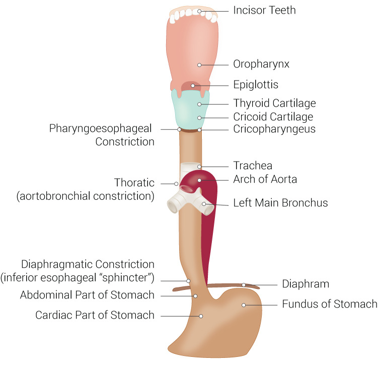
Anatomic constrictions of the oeophagus. During the course of the oesophagus, there are three anatomic constrictions, where the chance for a food impaction or foreign body is the most likely to occur. These sites are the pharyngo-oesophageal constriction, the aortobronchial constriction and the inferior oesophageal sphincter. English labels
Figure retrieved from Rosen RD, Winters R. Physiology, Lower Esophageal Sphincter. [Updated 2022 Apr 5]. In: StatPearls [Internet]. Treasure Island (FL): StatPearls Publishing; 2022 Jan- (CC BY).
Figure retrieved from Rosen RD, Winters R. Physiology, Lower Esophageal Sphincter. [Updated 2022 Apr 5]. In: StatPearls [Internet]. Treasure Island (FL): StatPearls Publishing; 2022 Jan- (CC BY).
Anatomical structures in item:
Uploaded by: rva
Netherlands, Leiden – Leiden University Medical Center, Leiden University
Dens incisivus
Pars oralis pharyngis
Oesophagus
Epiglottis
Cartilago thyroidea
Cartilago cricoidea
Musculus cricopharyngeus
Constrictio pharyngooesophagealis
Constrictio bronchoaortica oesophageae
Constrictio diaphragmatica oesophageae
Musculus sphincter cardiacus
Pars cardiaca gastricae
Fundus gastricus
Pars cardiaca gastricae
Diaphragma
Bronchus principalis sinister
Trachea
Carina tracheae
Arcus aortae
Creator(s)/credit: Beckie Palmer
Requirements for usage
You are free to use this item if you follow the requirements of the license:  View license
 View license
 If you use this item you should credit it as follows:
- For usage in print - copy and paste the line below:
- For digital usage (e.g. in PowerPoint, Impress, Word, Writer) - copy and paste the line below (optionally add the license icon):
"Palmer - Drawing Anatomic constrictions of the oeophagus - English labels" at AnatomyTOOL.org by Beckie Palmer, © StatPearls Publishing LLC, license: Creative Commons Attribution
"Palmer - Drawing Anatomic constrictions of the oeophagus - English labels" by Beckie Palmer, © StatPearls Publishing LLC, license: CC BY


Comments