nid: 63515
Additional formats:
None available
Description:
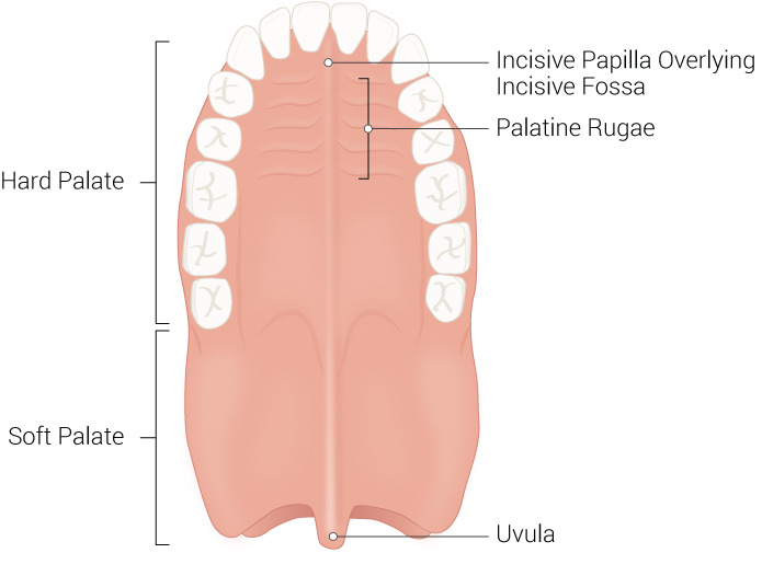
Palate and superior dental arch. The anterior two-thirds of the palate is the hard palate, which is formed by the maxilla and palatine bones. The soft palate is formed by muscles and connective tissue. English labels
Figure and description retrieved from Helwany M, Rathee M. Anatomy, Head and Neck, Palate. [Updated 2022 Jun 11]. In: StatPearls [Internet]. Treasure Island (FL): StatPearls Publishing; 2022 Jan- (CC BY).
Figure and description retrieved from Helwany M, Rathee M. Anatomy, Head and Neck, Palate. [Updated 2022 Jun 11]. In: StatPearls [Internet]. Treasure Island (FL): StatPearls Publishing; 2022 Jan- (CC BY).
Anatomical structures in item:
Uploaded by: rva
Netherlands, Leiden – Leiden University Medical Center, Leiden University
Palatum durum
Palatum molle
Palatum
Papilla incisiva
Fossa incisiva
Plicae palatinae transversae
Uvula palatina
Arcus dentalis maxillaris
Creator(s)/credit: Beckie Palmer
Requirements for usage
You are free to use this item if you follow the requirements of the license:  View license
 View license
 If you use this item you should credit it as follows:
- For usage in print - copy and paste the line below:
- For digital usage (e.g. in PowerPoint, Impress, Word, Writer) - copy and paste the line below (optionally add the license icon):
"Palmer - Drawing Palate and superior dental arch - English labels" at AnatomyTOOL.org by Beckie Palmer, © StatPearls Publishing LLC, license: Creative Commons Attribution
"Palmer - Drawing Palate and superior dental arch - English labels" by Beckie Palmer, © StatPearls Publishing LLC, license: CC BY


Comments