nid: 63500
Additional formats:
None available
Description:
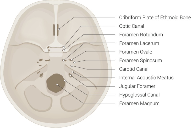
Foramina in base of skull. Illustration showing the cribriform plate of ethmoid bone, optic canal, foramen rotundum, foramen lacerum, foramen ovale, foramen spinosum, carotid canal, internal acoustic meatus, jugular foramer, hypoglossal canal and foramen magnum. English labels
Figure retrieved from Yu M, Wang SM. Anatomy, Head and Neck, Ethmoid Bone. [Updated 2022 Jul 25]. In: StatPearls [Internet]. Treasure Island (FL): StatPearls Publishing; 2022 Jan- (CC BY).
Figure retrieved from Yu M, Wang SM. Anatomy, Head and Neck, Ethmoid Bone. [Updated 2022 Jul 25]. In: StatPearls [Internet]. Treasure Island (FL): StatPearls Publishing; 2022 Jan- (CC BY).
Anatomical structures in item:
Uploaded by: rva
Netherlands, Leiden – Leiden University Medical Center, Leiden University
Basis cranii
Lamina cribrosa
Canalis opticus
Foramen rotundum
Foramen lacerum
Foramen ovale
Foramen spinosum
Canalis caroticus
Meatus acusticus internus
Foramen jugulare
Canalis nervi hypoglossi
Foramen magnum
Creator(s)/credit: Beckie Palmer
Requirements for usage
You are free to use this item if you follow the requirements of the license:  View license
 View license
 If you use this item you should credit it as follows:
- For usage in print - copy and paste the line below:
- For digital usage (e.g. in PowerPoint, Impress, Word, Writer) - copy and paste the line below (optionally add the license icon):
"Palmer - Drawing Foramina in base of skull - English labels" at AnatomyTOOL.org by Beckie Palmer, © StatPearls Publishing LLC, license: Creative Commons Attribution
"Palmer - Drawing Foramina in base of skull - English labels" by Beckie Palmer, © StatPearls Publishing LLC, license: CC BY


Comments