
nid: 63466
Additional formats:
None available
Description:
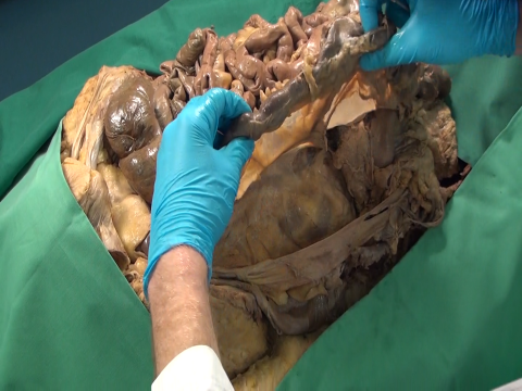
The video shows a human dissection specimen in which the descending colon and mesocolon are mobilized. In the embryological period they got attached to the back wall of the abdomen, leading to their so-called secondary retroperitoneal position. These attachments can be relatively easily detached. Such detachment procedures form the basis of abdominal surgery.
In this video, first the jejunum and ileum are laid aside. Then the mobilized descending colon and mesocolon are lifted. The blood vessels in the descending mesocolon can be seen. The parietal peritoneum of the body's side wall is shown. The area to which the descending colon and mesocolon lay attached, is pointed at. Before the embryological attachment the original back wall peritoneum lay here. Finally, the the mobilized descending colon and mesocolon are laid back to the location where they lay.
Anatomical structures in item:
Uploaded by: opgobee
Netherlands, Leiden – Leiden University Medical Center, Leiden University
Mesocolon descendens
Colon descendens
Peritoneum
Peritoneum parietale
Creator(s)/credit: Paul Gobée MD, anatomist, LUMC; Judith den Boeft, prosector, video, LUMC
Requirements for usage
You are free to use this item if you follow the requirements of the license:  View license
View license If you use this item you should credit it as follows:
- For usage in print - copy and paste the line below:
- For digital usage (e.g. in PowerPoint, Impress, Word, Writer) - copy and paste the line below (optionally add the license icon):
"Leiden - Video (Meso)colon descendens mobilized " at AnatomyTOOL.org by Paul Gobée, LUMC and Judith den Boeft, LUMC, license: Creative Commons Attribution-NonCommercial-ShareAlike
"Leiden - Video (Meso)colon descendens mobilized " by Paul Gobée, LUMC and Judith den Boeft, LUMC, license: CC BY-NC-SA


Comments