
nid: 63262
Additional formats:
None available
Description:
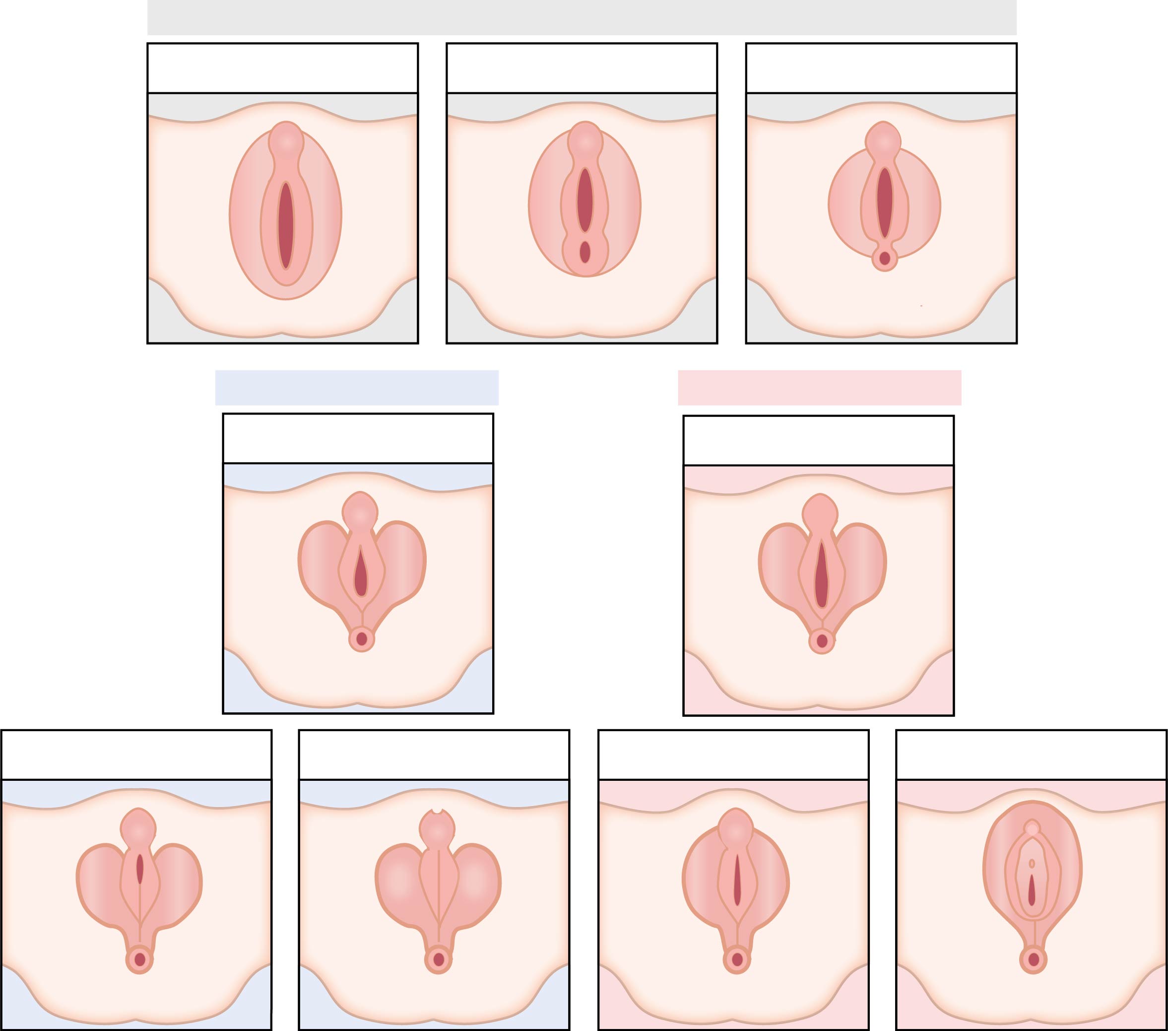
Differentiation of male and female external genitalia. The upper images depict the indifferent stage of the external genitalia in the 6th, 7th, and 9th week of embryonic development. In this stage, a separate anal orifice and urogenital orifice are formed. In the middle two images, the situation around the 10th week is drawn - the genitalia start to develop to either male with the forming of an early penis or female. Furthermore, the anus is formed and detached from the urethral groove by the perineum. The two lower left images show the differentiation of the male external genitalia in the 12th and 14th week, with the closing of the urethral groove and forming of scrotum and urinary meatus. The two lower right images show the differentiation of the female external genitalia in these weeks, characterized by the forming of the mons pubis, labia minora and majora, and later the clitoris, external urethral orifice and hymen. Version without labels.
Also published in Textbook of Obstetrics and Gynaecology, E.A.P. Stegers et al, 2019, BSL, https://doi.org/10.1007/978-90-368-2131-5, ISBN 978-90-368-2130-8
Also published in Textbook of Obstetrics and Gynaecology, E.A.P. Stegers et al, 2019, BSL, https://doi.org/10.1007/978-90-368-2131-5, ISBN 978-90-368-2130-8
Anatomical structures in item:
Uploaded by: rva
Netherlands, Leiden – Leiden University Medical Center, Leiden University
Organa genitalia feminina externa
Organa genitalia masculina externa
Organa genitalia feminina externa
Organa genitalia feminina externa
Perineum
Anus
Anus
Penis
Scrotum
Labium minus pudendi
Labium majus pudendi
Clitoris
Ostium urethrae externum (Urethra feminina)
Hymen
Mons pubis
Creator(s)/credit: Ron Slagter NZIMBI, medical illustrator; Prof. Marco C DeRuiter PhD, anatomist, professor of clinical and applied anatomy, LUMC
Requirements for usage
You are free to use this item if you follow the requirements of the license:  View license
View license If you use this item you should credit it as follows:
- For usage in print - copy and paste the line below:
- For digital usage (e.g. in PowerPoint, Impress, Word, Writer) - copy and paste the line below (optionally add the license icon):
"Leiden - Drawing Differentiation of male and female external genitalia - no labels" at AnatomyTOOL.org by Ron Slagter and Marco C DeRuiter, LUMC, license: Creative Commons Attribution-NonCommercial-ShareAlike


Comments