
nid: 63259
Additional formats:
None available
Description:
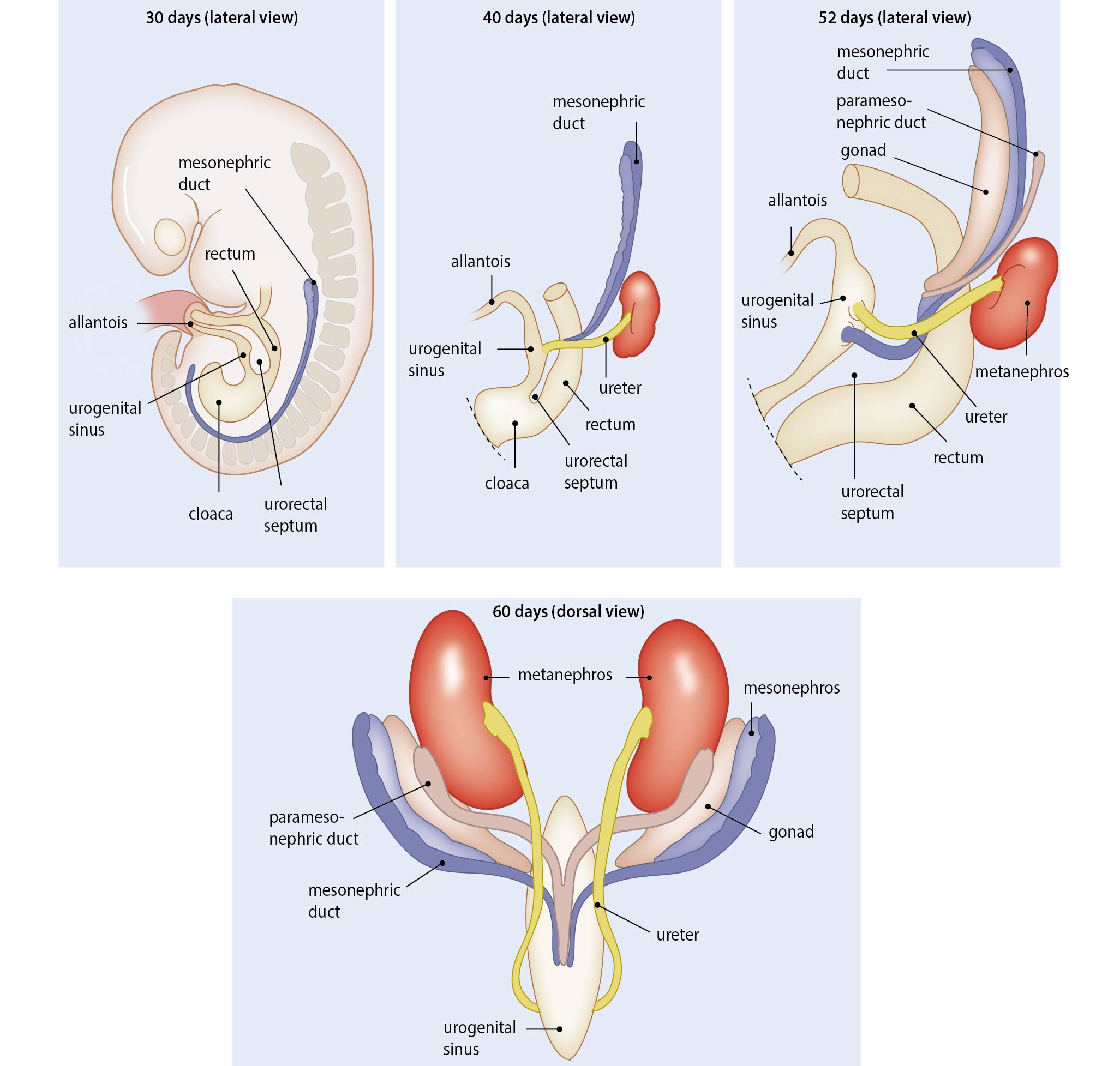
Embryonic development of the kidney. This image shows the development of the kidney around four different days, namely day 30, 40, 52 and 60. The relation between the mesonephric duct, paramesonephric duct, gonad and metanephros can be seen. English labels.
Also published in Textbook of Obstetrics and Gynaecology, E.A.P. Stegers et al, 2019, BSL, https://doi.org/10.1007/978-90-368-2131-5, ISBN 978-90-368-2130-8
Also published in Textbook of Obstetrics and Gynaecology, E.A.P. Stegers et al, 2019, BSL, https://doi.org/10.1007/978-90-368-2131-5, ISBN 978-90-368-2130-8
Anatomical structures in item:
Uploaded by: rva
Netherlands, Leiden – Leiden University Medical Center, Leiden University
Rectum
Ureter
Ren (Nephros)
Creator(s)/credit: Ron Slagter NZIMBI, medical illustrator; Bernadette S de Bakker MD, PhD, anatomist
Requirements for usage
You are free to use this item if you follow the requirements of the license:  View license
View license If you use this item you should credit it as follows:
- For usage in print - copy and paste the line below:
- For digital usage (e.g. in PowerPoint, Impress, Word, Writer) - copy and paste the line below (optionally add the license icon):
"Amsterdam - Drawing Embryonic development of the kidney - English labels" at AnatomyTOOL.org by Ron Slagter and Bernadette S de Bakker, license: Creative Commons Attribution-NonCommercial-ShareAlike


Comments