nid: 63252
Additional formats:
None available
Description:
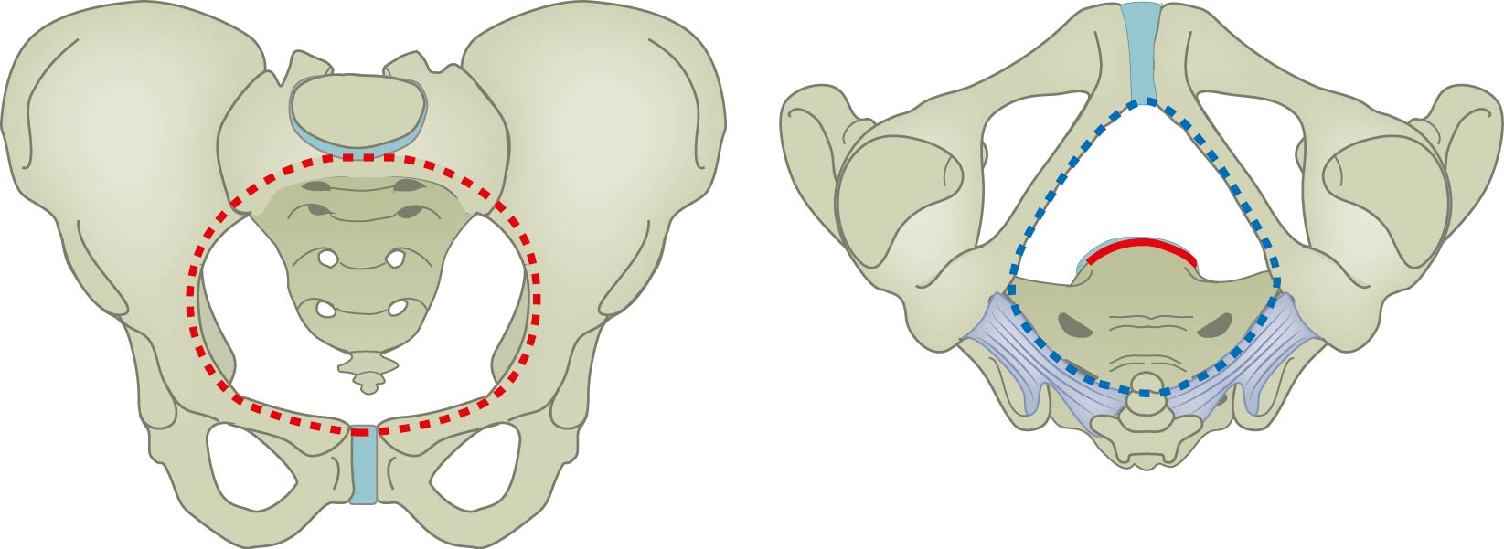
Pelvic inlet and outlet. Image showing the pelvic inlet in red (apertura pelvis superior) and pelvic outlet in blue (apertura pelvis inferior). The pelvic inlet is formed anteriorly by the pubic symphysis, posteriorly by the promontory of sacrum, and laterally by the ileopectineal (arcuate) lines. The pelvic outlet is also formed anteriorly by the symphysis pubis. However, its posterior border is the coccyx and its lateral borders are the isciopubic ramus (anterolateral) and sacrotuberous ligament (posterolateral). Version without labels.
Also published in Textbook of Obstetrics and Gynaecology, E.A.P. Stegers et al, 2019, BSL, https://doi.org/10.1007/978-90-368-2131-5, ISBN 978-90-368-2130-8
Also published in Textbook of Obstetrics and Gynaecology, E.A.P. Stegers et al, 2019, BSL, https://doi.org/10.1007/978-90-368-2131-5, ISBN 978-90-368-2130-8
Anatomical structures in item:
Uploaded by: rva
Netherlands, Leiden – Leiden University Medical Center, Leiden University
Apertura pelvis superior
Pelvis
Apertura pelvis inferior
Ilium
Articulatio sacroiliaca
Ischium
Tuber ischiadicum
Pubis
Symphysis pubica
Coccyx [vertebrae coccygeae I-IV]
Linea terminalis pelvis
Linea arcuata ilii
Arcuate line of hip bone
Promontorium ossis sacri
Os sacrum [vertebrae sacrales I - V]
Ramus ischiopubicus
Ligamentum sacrotuberale
Creator(s)/credit: Ron Slagter NZIMBI, medical illustrator; Prof. Marco C DeRuiter PhD, anatomist, professor of clinical and applied anatomy, LUMC
Requirements for usage
You are free to use this item if you follow the requirements of the license:  View license
 View license
 If you use this item you should credit it as follows:
- For usage in print - copy and paste the line below:
- For digital usage (e.g. in PowerPoint, Impress, Word, Writer) - copy and paste the line below (optionally add the license icon):
"Leiden - Drawing Pelvic inlet and outlet - no labels" at AnatomyTOOL.org by Ron Slagter and Marco C DeRuiter, LUMC, license: Creative Commons Attribution-NonCommercial-ShareAlike
"Leiden - Drawing Pelvic inlet and outlet - no labels" by Ron Slagter and Marco C DeRuiter, LUMC, license: CC BY-NC-SA


Comments