nid: 63189
Additional formats:
None available
Description:
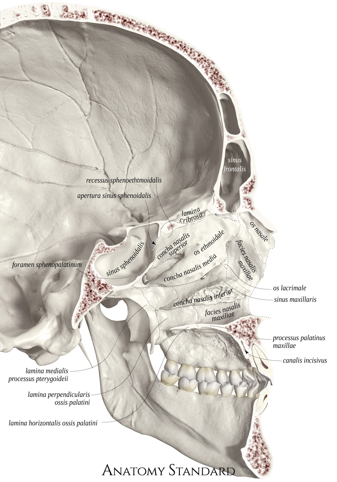
Lateral wall of nasal cavity: sagittal cut. The nasal cavity is the air-filled compartment above the palate, bordered by skeletal structures and cartilages, and covered from inside by mucosa. This parasagittal cut exposes the most structurally complex part of the nasal cavity: the lateral wall. The sphenoid sinus opens into the nasal cavity above the upper nasal concha — into the sphenoethmoid recess. Moreover, the pterygopalatine fossa communicates with the nasal cavity via the sphenopalatine foramen. The latter one localizes dorsally to the middle nasal concha. Latin labels.
Image and description retrieved from Anatomy Standard.
Image and description retrieved from Anatomy Standard.
Anatomical structures in item:
Uploaded by: rva
Netherlands, Leiden – Leiden University Medical Center, Leiden University
Cavitas nasi
Sinus frontalis
Sinus sphenoidalis
Recessus sphenoethmoidalis
Apertura sinus sphenoidalis
Lamina cribrosa
Foramen sphenopalatinum
Concha nasalis superior
Os ethmoidale
Os nasale
Facies nasalis corporis maxillae
Os lacrimale
Sinus maxillaris
Concha nasalis inferior
Concha nasalis media
Processus palatinus maxillae
Canales incisivus
Lamina horizontalis ossis palatini
Lamina perpendicularis ossis palatini
Lamina medialis processi pterygoideus ossis sphenoidalis
Creator(s)/credit: Jānis Šavlovskis MD, PhD, Assistant Professor; Kristaps Raits, 3D generalist
Requirements for usage
You are free to use this item if you follow the requirements of the license:  View license
 View license
 If you use this item you should credit it as follows:
- For usage in print - copy and paste the line below:
- For digital usage (e.g. in PowerPoint, Impress, Word, Writer) - copy and paste the line below (optionally add the license icon):
"Anatomy Standard - Drawing Lateral wall of nasal cavity: sagittal cut - Latin labels" at AnatomyTOOL.org by Jānis Šavlovskis and Kristaps Raits, license: Creative Commons Attribution-NonCommercial


Comments