
nid: 63093
Additional formats:
None available
Description:
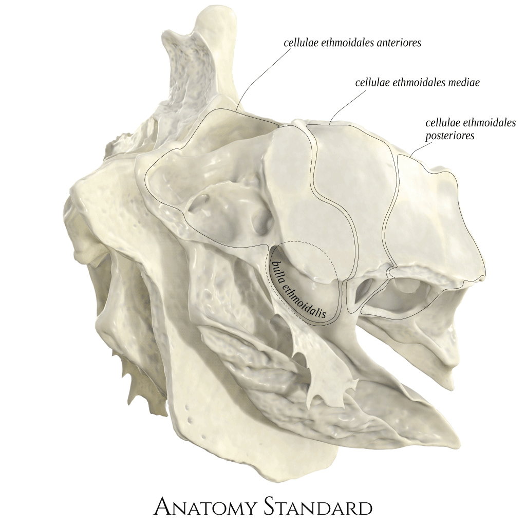
Ethnoid bone: inferior ventral view. The ethmoid bone is localized between the orbits and is a significant part of the nasal cavity. It is an unpaired bone, made almost entirely by thin bony lamellae. The inferior ventral view of ethmoid bone demonstrates multiple exposed cellulae ethmoidales anteriores that in situ are covered by the frontal bone with frontal sinus from the top and with lacrimal bone from aside. Version without labels.
Image and description retrieved from Anatomy Standard. Via this link more images can be found, including oblique views.
Image and description retrieved from Anatomy Standard. Via this link more images can be found, including oblique views.
Anatomical structures in item:
Uploaded by: rva
Netherlands, Leiden – Leiden University Medical Center, Leiden University
Os ethmoidale
Bulla ethmoidalis
Cellulae ethmoidales
Cellulae ethmoidales anteriores
Cellulae ethmoidales mediae
Cellulae ethmoidales posteriores
Creator(s)/credit: Jānis Šavlovskis MD, PhD, Assistant Professor; Kristaps Raits, 3D generalist
Requirements for usage
You are free to use this item if you follow the requirements of the license:  View license
View license If you use this item you should credit it as follows:
- For usage in print - copy and paste the line below:
- For digital usage (e.g. in PowerPoint, Impress, Word, Writer) - copy and paste the line below (optionally add the license icon):
"Anatomy Standard - Drawing Ethmoid bone: inferior ventral view - no labels" at AnatomyTOOL.org by Jānis Šavlovskis and Kristaps Raits, license: Creative Commons Attribution-NonCommercial


Comments