nid: 62958
Additional formats:
None available
Description:
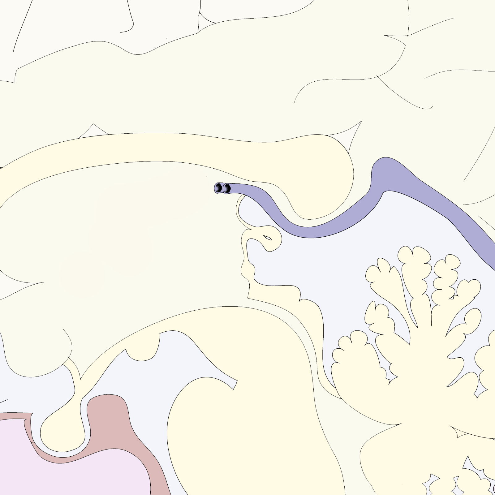
Anatomy of the pineal gland region. The pineal gland is located between the thalamic bodies. The gland produces melatonin, which influences sleep and seasonal patterns. Version without labels.
Case courtesy of Assoc Prof Frank Gaillard, Radiopaedia.org. From the case rID: 10766
Case courtesy of Assoc Prof Frank Gaillard, Radiopaedia.org. From the case rID: 10766
Anatomical structures in item:
Uploaded by: rva
Netherlands, Leiden – Leiden University Medical Center, Leiden University
Corpus callosum
Vena magna cerebri
Habenula
Corpus pineale
Recessus pinealis
Commissura posterior
Colliculus superior
Aqueductus mesencephali
Mesencephalon
Glandula pituitaria
Hypothalamus
Creator(s)/credit: Dr Frank Gaillard MB.BS, MMed
Requirements for usage
You are free to use this item if you follow the requirements of the license:  View license
 View license
 If you use this item you should credit it as follows:
- For usage in print - copy and paste the line below:
- For digital usage (e.g. in PowerPoint, Impress, Word, Writer) - copy and paste the line below (optionally add the license icon):
"Radiopaedia - Drawing Anatomy of the pineal gland region - no labels" at AnatomyTOOL.org by Frank Gaillard, license: Creative Commons Attribution-NonCommercial-ShareAlike


Comments