nid: 62689
Additional formats:
None available
Description:
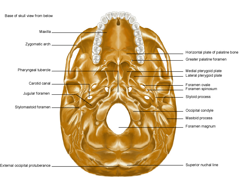
External surface of cranial base. Drawing of the anatomy of the cranial base. The cranial foramina are shown, through which the cranial nerves travel. English labels.
This image by the Royal College of Surgeons of Ireland (RCSI) is retrieved from Health Education Assets Library (HEAL) of the University of Utah.
This image by the Royal College of Surgeons of Ireland (RCSI) is retrieved from Health Education Assets Library (HEAL) of the University of Utah.
Anatomical structures in item:
Uploaded by: rva
Netherlands, Leiden – Leiden University Medical Center, Leiden University
Basis cranii
Maxilla
Arcus zygomaticus
Tuberculum pharyngeum
Canalis caroticus
Foramen jugulare
Foramen stylomastoideum
Protuberantia occipitalis externa
Linea nuchalis superior
Foramen magnum
Processus mastoideus
Condylus occipitalis
Processus styloideus
Foramen spinosum
Foramen ovale
Foramen palatinum majus
Lamina horizontalis ossis palatini
Dentes
Creator(s)/credit: Royal College of Surgeons of Ireland
Requirements for usage
You are free to use this item if you follow the requirements of the license:  View license
 View license
 If you use this item you should credit it as follows:
- For usage in print - copy and paste the line below:
- For digital usage (e.g. in PowerPoint, Impress, Word, Writer) - copy and paste the line below (optionally add the license icon):
"RCSI - Drawing External surface of cranial base - English labels" at AnatomyTOOL.org by Royal College of Surgeons of Ireland, license: Creative Commons Attribution-NonCommercial-ShareAlike


Comments