nid: 62688
Additional formats:
None available
Description:
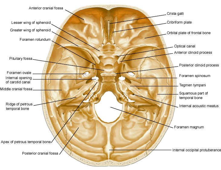
Internal surface of cranial base. Drawing of the anatomy of the cranial base. The cranial foramina are shown, through which the cranial nerves travel. English labels.
This image by the Royal College of Surgeons of Ireland (RCSI) is retrieved from Health Education Assets Library (HEAL) of the University of Utah.
This image by the Royal College of Surgeons of Ireland (RCSI) is retrieved from Health Education Assets Library (HEAL) of the University of Utah.
Anatomical structures in item:
Uploaded by: rva
Netherlands, Leiden – Leiden University Medical Center, Leiden University
Basis cranii
Fossa cranii anterior
Ala minor ossis sphenoidalis
Ala major ossis sphenoidalis
Os sphenoidale
Foramen rotundum
Fossa hypophysialis
Foramen ovale
Apertura interna canalis carotici
Canalis caroticus
Fossa cranii media
Pars petrosa ossis temporalis
Apex partis petrosae ossis temporalis
Fossa cranii posterior
Protuberantia occipitalis interna
Foramen magnum
Porus acusticus internus
Pars squamosa ossis temporalis
Tegmen tympani
Foramen spinosum
Processus clinoideus posterior
Processus clinoideus anterior
Canalis opticus
Lamina cribrosa
Crista galli
Creator(s)/credit: Royal College of Surgeons of Ireland
Requirements for usage
You are free to use this item if you follow the requirements of the license:  View license
 View license
 If you use this item you should credit it as follows:
- For usage in print - copy and paste the line below:
- For digital usage (e.g. in PowerPoint, Impress, Word, Writer) - copy and paste the line below (optionally add the license icon):
"RCSI - Drawing Internal surface of cranial base - English labels" at AnatomyTOOL.org by Royal College of Surgeons of Ireland, license: Creative Commons Attribution-NonCommercial-ShareAlike


Comments