
nid: 62622
Additional formats:
None available
Description:
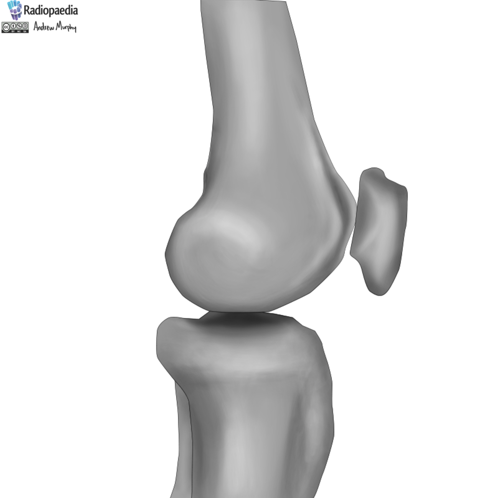
Bones of the knee joint: medial view. This drawing shows the different parts of the bones of the knee.
Case courtesy of Dr Henry Knipe, Radiopaedia.org. From the case rID: 31397
Case courtesy of Dr Henry Knipe, Radiopaedia.org. From the case rID: 31397
Anatomical structures in item:
Uploaded by: rva
Netherlands, Leiden – Leiden University Medical Center, Leiden University
Genu
Articulatio genus
Femur
Epicondylus medialis femoris
Fossa intercondylaris
Condylus medialis femoris
Condylus medialis tibiae
Tibia
Tuberositas tibiae
Fibula
Facies patellaris femoris
Basis patellae
Area intercondylaris anterior
Area intercondylaris posterior
Tuberculum adductorium femoris
Creator(s)/credit: Dr Andrew Murphy
Requirements for usage
You are free to use this item if you follow the requirements of the license:  View license
View license If you use this item you should credit it as follows:
- For usage in print - copy and paste the line below:
- For digital usage (e.g. in PowerPoint, Impress, Word, Writer) - copy and paste the line below (optionally add the license icon):
"Radiopaedia - Drawing Bones of the knee joint: medial view - no labels" at AnatomyTOOL.org by Andrew Murphy, license: Creative Commons Attribution-NonCommercial-ShareAlike


Comments