
nid: 62612
Additional formats:
None available
Description:
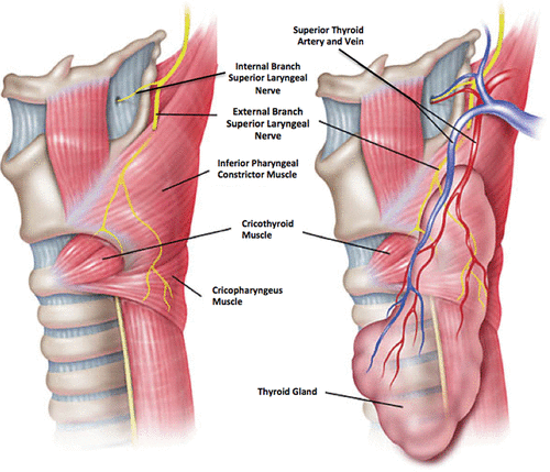
Lateral view of larynx: superior laryngeal nerve (SLN). The SLN arises from the nodose ganglion as a branch of the vagal nerve within the carotid sheath midway between the jugular foramen and the carotid bifurcation (at the level of the C2 vertebra). It then runs medially and caudally approximately 1.5 cm toward the thyrohyoid membrane before dividing into the internal and external branches.
Image and description source: Tempel ZJ, Smith JS, Shaffrey C, Arnold PM, Fehlings MG, Mroz TE, Riew KD, Kanter AS. A Multicenter Review of Superior Laryngeal Nerve Injury Following Anterior Cervical Spine Surgery. Global Spine J. 2017 Apr;7(1 Suppl):7S-11S (CC BY-NC-ND).
Image and description source: Tempel ZJ, Smith JS, Shaffrey C, Arnold PM, Fehlings MG, Mroz TE, Riew KD, Kanter AS. A Multicenter Review of Superior Laryngeal Nerve Injury Following Anterior Cervical Spine Surgery. Global Spine J. 2017 Apr;7(1 Suppl):7S-11S (CC BY-NC-ND).
Anatomical structures in item:
Uploaded by: rva
Netherlands, Leiden – Leiden University Medical Center, Leiden University
Larynx
Ramus internus nervus laryngei superioris
Nervus laryngeus superior
Ramus externus nervus laryngei superioris
Musculus constrictor pharyngis inferior
Musculus cricothyroideus
Musculus cricopharyngeus
Glandula thyroidea
Arteria thyroidea superior
Vena thyroidea superior
Creator(s)/credit: ZJ Tempel; JS Smith; C Shaffrey; PM Arnold; MG Fehlings; TE Mroz; KD Riew; AS Kanter
Requirements for usage
You are free to use this item if you follow the requirements of the license:  View license
View license If you use this item you should credit it as follows:
- For usage in print - copy and paste the line below:
- For digital usage (e.g. in PowerPoint, Impress, Word, Writer) - copy and paste the line below (optionally add the license icon):
"Tempel - Drawing Lateral view of larynx: superior laryngeal nerve - English labels" at AnatomyTOOL.org by ZJ Tempel, JS Smith, C Shaffrey et al, license: Creative Commons Attribution-NonCommercial-NoDerivs
"Tempel - Drawing Lateral view of larynx: superior laryngeal nerve - English labels" by ZJ Tempel, JS Smith, C Shaffrey et al, license: CC BY-NC-ND


Comments