nid: 62550
Additional formats:
None available
Description:
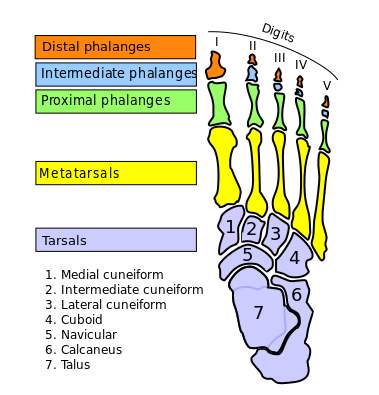
Bones of the foot. This image shows the different bones of the foot, namely the different tarsal, metatarsal and phalanx bones. English labels
This image was retrieved from Wikimedia Commons. This image is a derivative of the original by Mario Modesto Mata, it was edited by Johnuniq and HLHJ.
Anatomical structures in item:
Uploaded by: rva
Netherlands, Leiden – Leiden University Medical Center, Leiden University
Pes
Regio pedis
Digiti pedis
Hallux
Digitus secundus [II] pedis
Digitus tertius [III] pedis
Digitus quartus [IV] pedis
Digitus minimus pedis
Phalanx distalis pedis
Phalanx media pedis
Phalanx proximalis pedis
Ossa metatarsalia [I-V]
Ossa tarsalia
Os cuneiforme mediale
Os cuneiforme intermedium
Os cuneiforme laterale
Os cuboideum
Os naviculare
Calcaneus
Talus
Creator(s)/credit: Mario Modesto Mata; Johnuniq; HLHJ
Requirements for usage
You are free to use this item if you follow the requirements of the license:  View license
 View license
 If you use this item you should credit it as follows:
- For usage in print - copy and paste the line below:
- For digital usage (e.g. in PowerPoint, Impress, Word, Writer) - copy and paste the line below (optionally add the license icon):
"Modesto Mata - Drawing Bones of the foot - English labels" at AnatomyTOOL.org by Mario Modesto Mata, Johnuniq and HLHJ, license: Creative Commons Attribution-ShareAlike


Comments