
nid: 62397
Additional formats:
None available
Description:
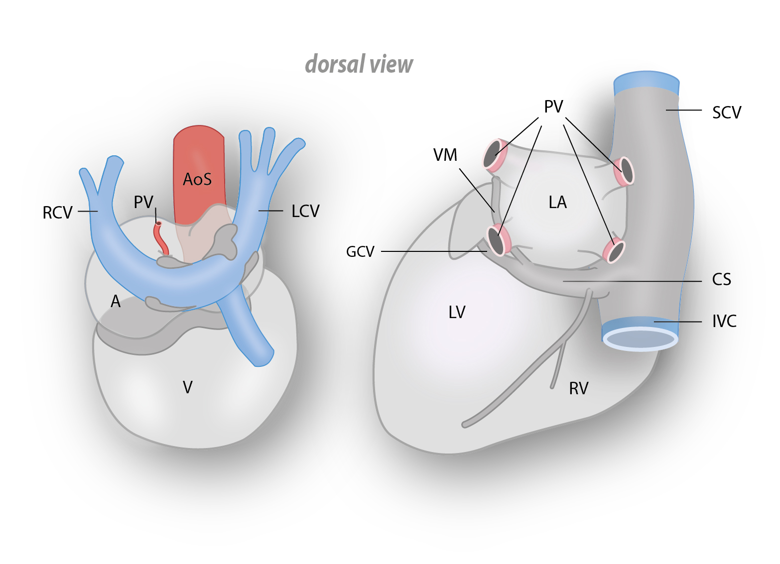
Dorsal view of embryonal and adult heart. The left drawing shows the dorsal side of the embryonal heart and the right drawing shows the dorsal side of the adult heart. Abbreviations: A: common atrium, AoS: aortic sac, CS: coronary sinus, GCV: great cardiac vein, ICV: inferior caval vein, LA: left atrium, LCV: left cardinal vein, LV: (putative) left ventricle, PV: pulmonary vein(s), RCV: right cardinal vein, RV: (putative) right ventricle, SCV: superior caval vein, V: common ventricle, VM: vein of Marshall/ Marshall ligament, English labels.
Source: Fig. 9. Working model for the developmental background of clinical arrhythmias in the child/adult. from Jongbloed MR, Vicente Steijn R, Hahurij ND, Kelder TP, Schalij MJ, Gittenberger-de Groot AC, Blom NA. Normal and abnormal development of the cardiac conduction system; implications for conduction and rhythm disorders in the child and adult. Differentiation. 2012 Jul;84(1):131-48.
Source: Fig. 9. Working model for the developmental background of clinical arrhythmias in the child/adult. from Jongbloed MR, Vicente Steijn R, Hahurij ND, Kelder TP, Schalij MJ, Gittenberger-de Groot AC, Blom NA. Normal and abnormal development of the cardiac conduction system; implications for conduction and rhythm disorders in the child and adult. Differentiation. 2012 Jul;84(1):131-48.
Anatomical structures in item:
Uploaded by: rva
Netherlands, Leiden – Leiden University Medical Center, Leiden University
Cor
Sinus coronarius
Vena cardiaca magna
Vena cava inferior
Atrium sinistrum
Ligamentum cardinale
Ventriculus sinister
Venae pulmonales
Ventriculus dexter
Vena cava superior
Vena obliqua atrii sinistri
Creator(s)/credit: Ron Slagter NZIMBI, medical illustrator; Dr Monique R.M. Jongbloed PhD, cardiologist, anatomist, LUMC
Requirements for usage
You are free to use this item if you follow the requirements of the license:  View license
View license If you use this item you should credit it as follows:
- For usage in print - copy and paste the line below:
- For digital usage (e.g. in PowerPoint, Impress, Word, Writer) - copy and paste the line below (optionally add the license icon):
"Leiden - Drawing Dorsal view of embryonal and adult heart - English labels" at AnatomyTOOL.org by Ron Slagter and Monique R.M. Jongbloed, LUMC, © Elsevier, license: Creative Commons Attribution-NonCommercial-ShareAlike
"Leiden - Drawing Dorsal view of embryonal and adult heart - English labels" by Ron Slagter and Monique R.M. Jongbloed, LUMC, © Elsevier, license: CC BY-NC-SA


Comments