
nid: 62316
Additional formats:
None available
Description:
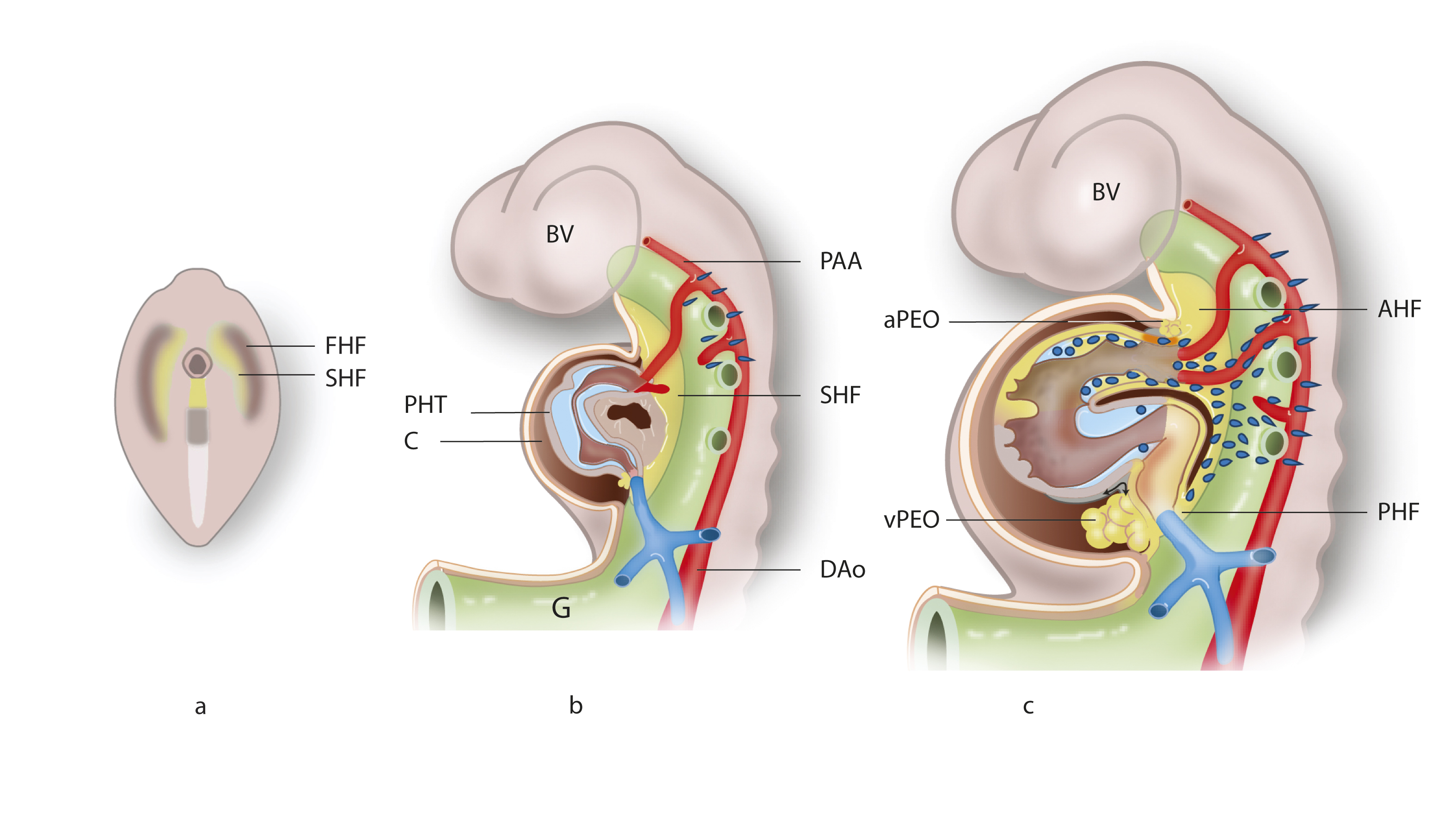
Development of the heart from the first and second heart fields. a) The cardiac crescent contains both first heart field (FHF) and second heart field (SHF) precursors. b) In a later stage, the still undivided heart with an atrial component with the cardinal veins and the pulmonary vein. The ventricular compartment shows anterior (future right) and posterior (future left) sides. The cardiac jelly has developed into two cushion complexes: the atrioventricular and outflow tract cushions (blue). The arterial pole shows the aortic sac and with the attached pharyngeal arch arteries. The dorsal mesocardium starts to disrupt (brown hole). The second heart field is depicted in yellow. c) The derivatives of the anterior and posterior parts of the second heart field (yellow) together with the migrated neural crest cells (blue-green). DAo, dorsal aorta; FHF, first heart field; PAA, pharyngeal arch arteries; SHF, second heart field. Description adapted from Poelmann RE, Gittenberger-de Groot AC. Development and evolution of the metazoan heart. Dev Dyn. 2019 Aug;248(8):634-656 (CC BY). English labels
Source: Fig. 2. Development of the heart from the first (FHF) and second (SHF) heart fields from Jongbloed MR, Vicente Steijn R, Hahurij ND, Kelder TP, Schalij MJ, Gittenberger-de Groot AC, Blom NA. Normal and abnormal development of the cardiac conduction system; implications for conduction and rhythm disorders in the child and adult. Differentiation. 2012 Jul;84(1):131-48.
Source: Fig. 2. Development of the heart from the first (FHF) and second (SHF) heart fields from Jongbloed MR, Vicente Steijn R, Hahurij ND, Kelder TP, Schalij MJ, Gittenberger-de Groot AC, Blom NA. Normal and abnormal development of the cardiac conduction system; implications for conduction and rhythm disorders in the child and adult. Differentiation. 2012 Jul;84(1):131-48.
Anatomical structures in item:
Uploaded by: rva
Netherlands, Leiden – Leiden University Medical Center, Leiden University
Cor
Ramus pharyngeus ateria palatinae descendentis
Aorta
Creator(s)/credit: Ron Slagter NZIMBI, medical illustrator; prof Adri C. Gittenberger-de Groot PhD, anatomist, head of dept. anatomy & embryology, LUMC; prof Robbert E. Poelmann PhD, anatomist, LUMC; Prof. Marco C. DeRuiter PhD, anatomist, professor of clinical and applied anatomy, LUMC
Requirements for usage
You are free to use this item if you follow the requirements of the license:  View license
View license If you use this item you should credit it as follows:
- For usage in print - copy and paste the line below:
- For digital usage (e.g. in PowerPoint, Impress, Word, Writer) - copy and paste the line below (optionally add the license icon):
"Leiden - Drawing Development of the heart from the first and second heart fields - English labels" at AnatomyTOOL.org by Ron Slagter, Adri C. Gittenberger-de Groot, LUMC, Robbert E. Poelmann, LUMC et al, © Elsevier, license: Creative Commons Attribution-NonCommercial-ShareAlike
"Leiden - Drawing Development of the heart from the first and second heart fields - English labels" by Ron Slagter, Adri C. Gittenberger-de Groot, LUMC, Robbert E. Poelmann, LUMC et al, © Elsevier, license: CC BY-NC-SA


Comments