
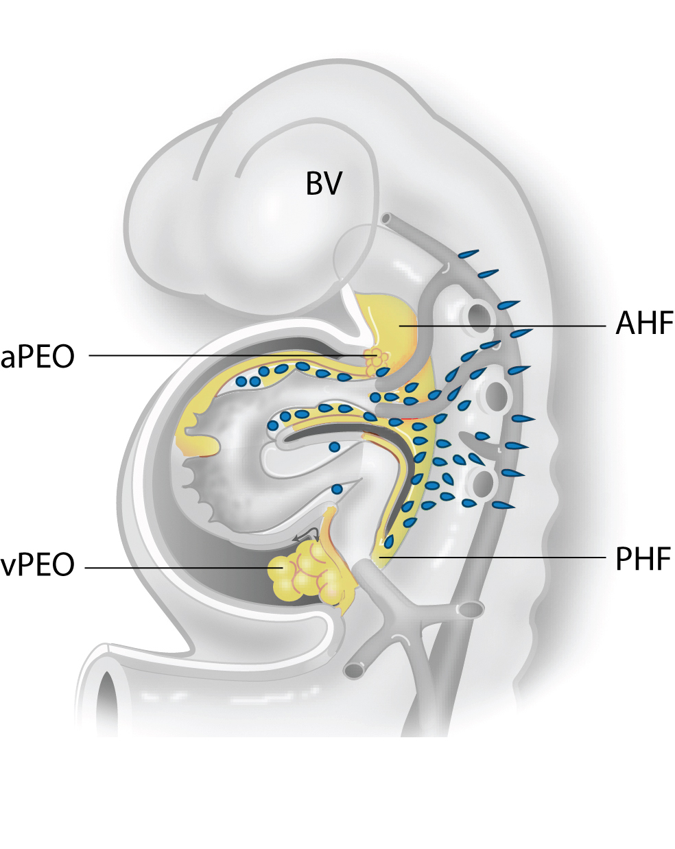
Schematic overview of the developing heart tube. "The second heart field is indicated contributing to the arterial pole of the heart including the great vessels and the right ventricle by the anterior population (AHF). At the venous pole SHF cells are entering from the posterior population (PHF). Both at the venous (vPEO) and arterial (aPEO) pole a proepicardial organ provides the epicardial cells that cover the myocardium and the intrapericardiac part of the great vessels. Neural crest cells migrate from the neural tube primarily to the arterial pole of the heart." Description from Fig. 1 of Grewal N, DeRuiter MC, Jongbloed MR, Goumans MJ, Klautz RJ, Poelmann RE, Gittenberger-de Groot AC. Normal and abnormal development of the aortic wall and valve: correlation with clinical entities. Neth Heart J. 2014 Sep;22(9):363-9 (CC BY).
 View license
 If you use this item you should credit it as follows:
- For usage in print - copy and paste the line below:
- For digital usage (e.g. in PowerPoint, Impress, Word, Writer) - copy and paste the line below (optionally add the license icon):


Comments