nid: 62220
Additional formats:
None available
Description:
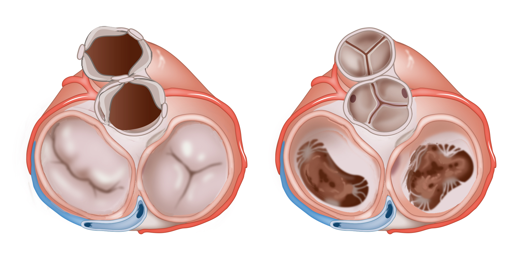
Valves of the heart during diastole and systole: superior view. During diastole (right), the two atrioventricular valves are open and the two semilunar valves are closed. During systole (left), the two atrioventricular valves are closed and the two semilunar valves are open.
Anatomical structures in item:
Uploaded by: rva
Netherlands, Leiden – Leiden University Medical Center, Leiden University
Cor
Valva mitralis
Valva tricuspidalis
Valva aortae
Valva trunci pulmonalis
Sinus coronarius
Arteria coronaria dextra
Arteria coronaria sinistra
Musculus papillaris
Creator(s)/credit: Ron Slagter NZIMBI, medical illustrator
Requirements for usage
You are free to use this item if you follow the requirements of the license:  View license
 View license
 If you use this item you should credit it as follows:
- For usage in print - copy and paste the line below:
- For digital usage (e.g. in PowerPoint, Impress, Word, Writer) - copy and paste the line below (optionally add the license icon):
"Slagter - Drawing Valves of the heart during diastole and systole: superior view - no labels" at AnatomyTOOL.org by Ron Slagter, license: Creative Commons Attribution-NonCommercial-ShareAlike


Comments