nid: 62210
Additional formats:
None available
Description:
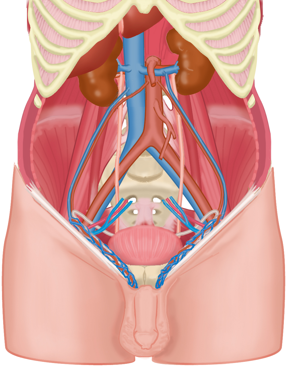
Layers of the abdominal wall: posterior abdominal wall. An image in a series, in which layers of the abdominal wall are consecutively removed. Originally created for e-learning 'CASK Inguinal Area' at https://www.caskanatomy.info/inguinalarea
Anatomical structures in item:
Uploaded by: rva
Netherlands, Leiden – Leiden University Medical Center, Leiden University
Abdomen
Ren (Nephros)
Aorta
Aorta abdominalis
Vena cava inferior
Truncus coeliacus
Arteria testicularis
Arcus costalis
Vesica urinaria
Ductus deferens
Ureter
Vena renalis dextra
Vena renalis sinistra
Venae renales
Arteria renalis
Arteria iliaca communis
Vena iliaca communis
Musculus psoas major
Musculus iliacus
Musculus piriformis
Funiculus spermaticus
Creator(s)/credit: Ron Slagter NZIMBI, medical illustrator; O. Paul Gobée MD, anatomist, LUMC
Requirements for usage
You are free to use this item if you follow the requirements of the license:  View license
 View license
 If you use this item you should credit it as follows:
- For usage in print - copy and paste the line below:
- For digital usage (e.g. in PowerPoint, Impress, Word, Writer) - copy and paste the line below (optionally add the license icon):
"Leiden - Drawing Layers of the abdominal wall: posterior abdominal wall - no labels" at AnatomyTOOL.org by Ron Slagter and O. Paul Gobée, LUMC, license: Creative Commons Attribution-NonCommercial-ShareAlike


Comments