
nid: 62203
Additional formats:
None available
Description:
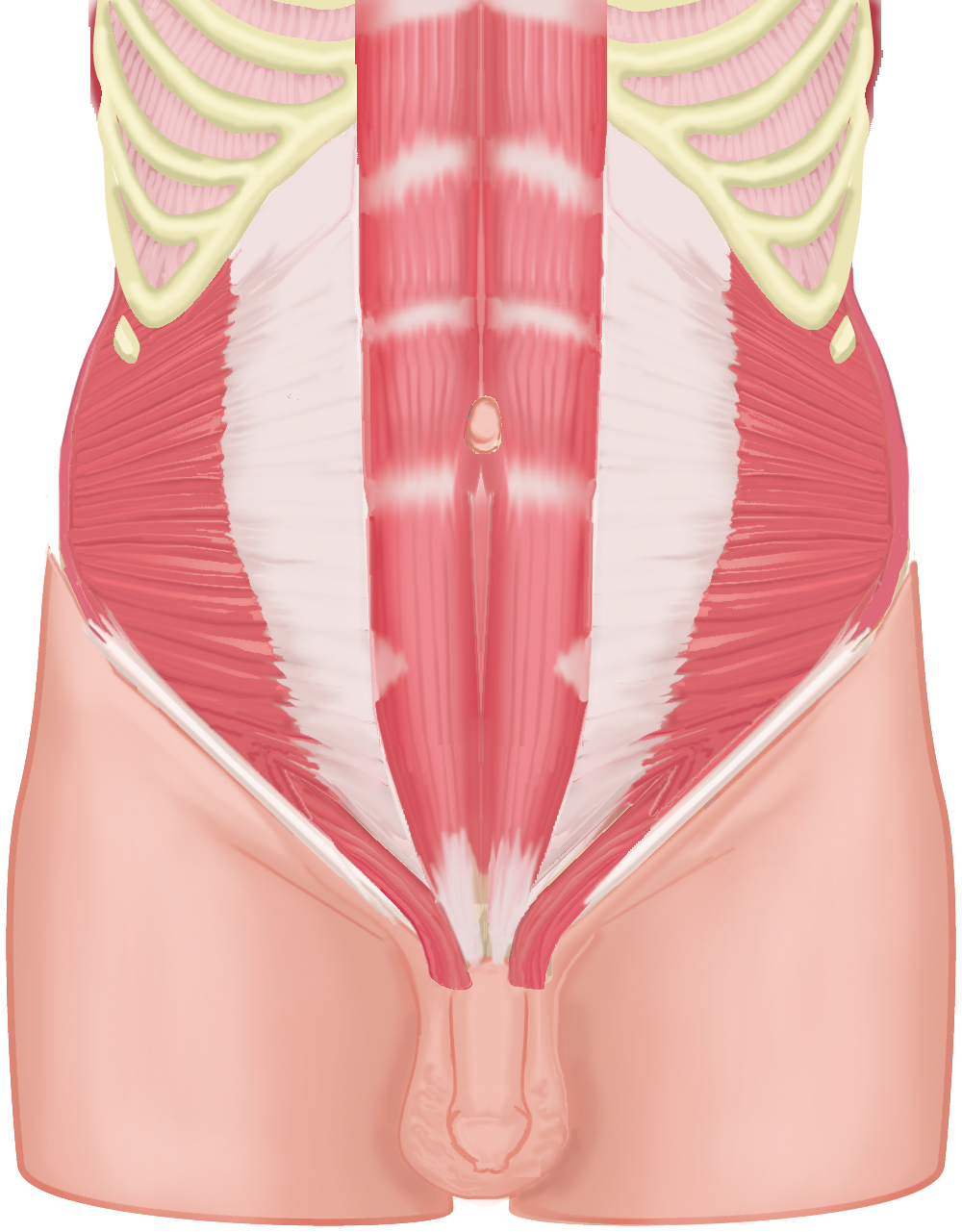
Layers of the abdominal wall: rectus abdominis muscle. An image in a series, in which layers of the abdominal wall are consecutively removed. Originally created for e-learning 'CASK Inguinal Area' at https://www.caskanatomy.info/inguinalarea
Anatomical structures in item:
Uploaded by: rva
Netherlands, Leiden – Leiden University Medical Center, Leiden University
Abdomen
Musculus rectus abdominis
Arcus costalis
Musculus cremaster
Ligamentum inguinale
Musculus obliquus internus abdominis
Creator(s)/credit: Ron Slagter NZIMBI, medical illustrator; O. Paul Gobée MD, anatomist, LUMC
Requirements for usage
You are free to use this item if you follow the requirements of the license:  View license
View license If you use this item you should credit it as follows:
- For usage in print - copy and paste the line below:
- For digital usage (e.g. in PowerPoint, Impress, Word, Writer) - copy and paste the line below (optionally add the license icon):
"Leiden - Drawing Layers of the abdominal wall: rectus abdominis muscle - no labels" at AnatomyTOOL.org by Ron Slagter and O. Paul Gobée, LUMC, license: Creative Commons Attribution-NonCommercial-ShareAlike


Comments