
nid: 62135
Additional formats:
None available
Description:
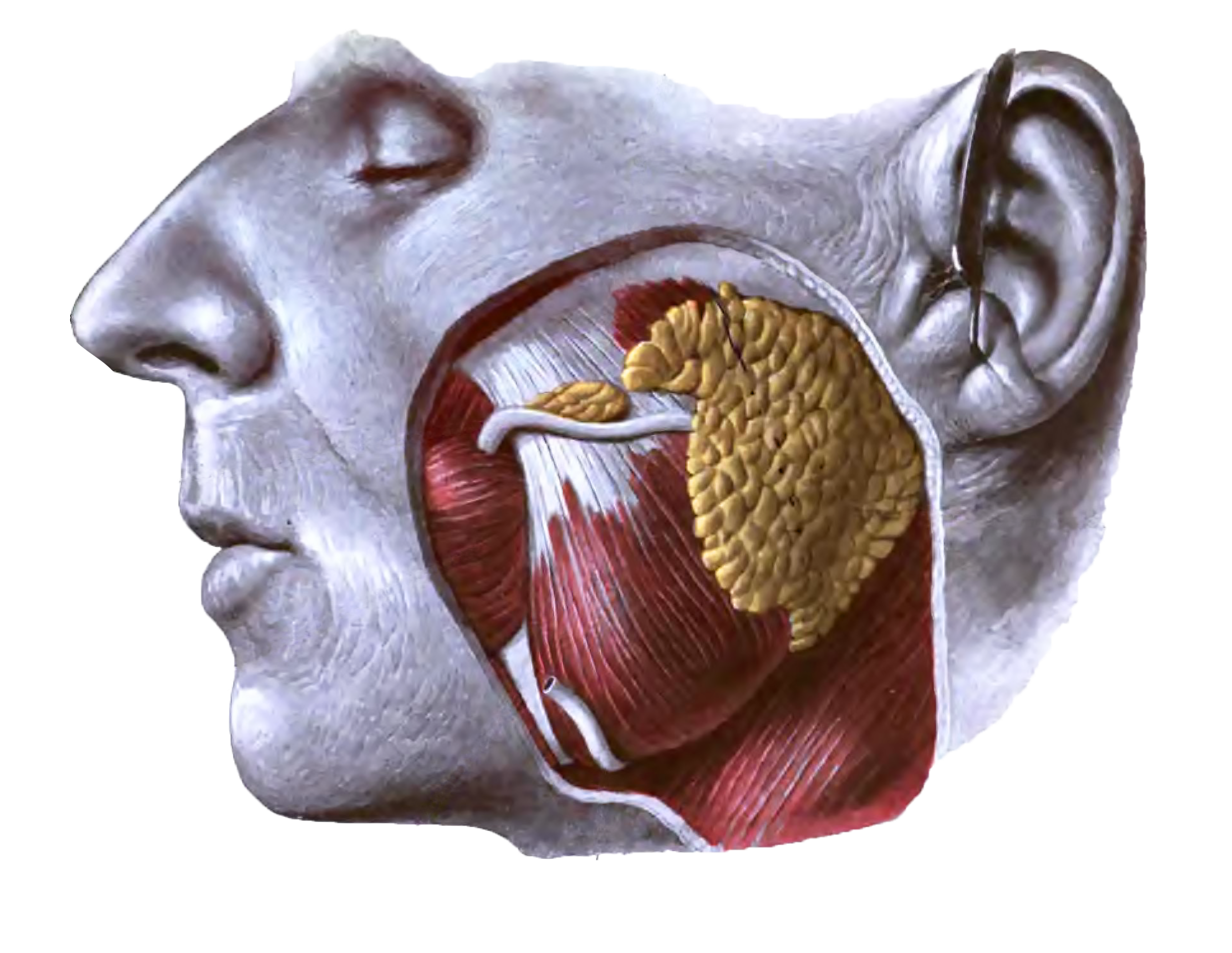
The parotid gland: view of the gland in position. Lateral view.
From 'Atlas and Textbook of Human Anatomy', 1906, Vol. 2, fig.358, by Johannes Sobotta and J. Playfair McMurrich. Artist: K. Hajek. Retrieved from Sobotta's Anatomy plates at Wikimedia. Possible original source: Sobotta's atlas at archive.org.
Image editing by dream_studio3.
From 'Atlas and Textbook of Human Anatomy', 1906, Vol. 2, fig.358, by Johannes Sobotta and J. Playfair McMurrich. Artist: K. Hajek. Retrieved from Sobotta's Anatomy plates at Wikimedia. Possible original source: Sobotta's atlas at archive.org.
Image editing by dream_studio3.
Anatomical structures in item:
Uploaded by: rva
Netherlands, Leiden – Leiden University Medical Center, Leiden University
Glandula parotidea
Musculus masseter
Musculus buccinator
Ductus parotideus
Arteria facialis
Glandulae parathyroideae accessoriae
Creator(s)/credit: Prof.dr. Johannes Sobotta, anatomist; dream_studio3 BA, image editing
Requirements for usage
You are free to use this item if you follow the requirements of the license:  View license
View license If you use this item you should credit it as follows:
- For usage in print - copy and paste the line below:
- For digital usage (e.g. in PowerPoint, Impress, Word, Writer) - copy and paste the line below (optionally add the license icon):
"Sobotta 1906 fig.358 - The parotid gland - no labels" at AnatomyTOOL.org by Johannes Sobotta and dream_studio3, license: Creative Commons Attribution-ShareAlike


Comments