nid: 62111
Additional formats:
None available
Description:
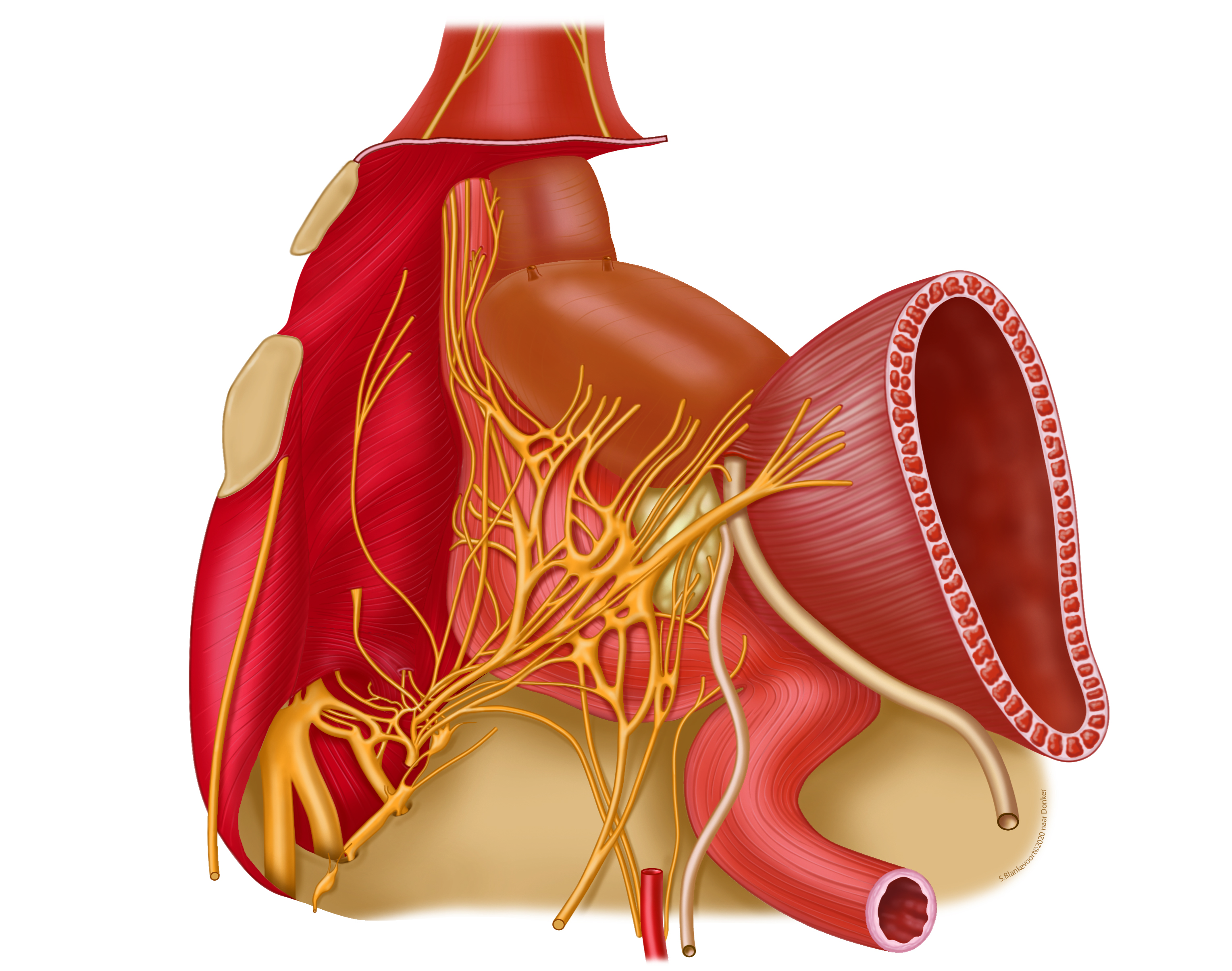
Neuroanatomy of the pelvic plexus. A dissection of the left pelvic plexus (inferior hypogastric plexus). The urogenitalia have been retracted to the right side. The peritoneum, pelvic vessels, pelvic fascia and pubic symphysis have been removed.
Coloured illustration based on the black-white drawings of prof dr P.J. Donker in 1982 (LUMC). A more thorough description can be found in J Urol, 128: 492-7, 1982.
Coloured illustration based on the black-white drawings of prof dr P.J. Donker in 1982 (LUMC). A more thorough description can be found in J Urol, 128: 492-7, 1982.
Anatomical structures in item:
Uploaded by: rva
Netherlands, Leiden – Leiden University Medical Center, Leiden University
Vesica urinaria
Plexus nervosus hypogastricus inferior
Plexus nervosus vesicalis
Plexus nervosus rectalis superior
Prostata (Glandula prostatica)
Ureter
Rectum
Truncus sympathicus
Nervus hypogastricus
Vesicula seminalis
Creator(s)/credit: Bas (S) Blankevoort, biology and medical illustrator, LUMC; Prof. Marco C DeRuiter PhD, anatomist, professor of clinical and applied anatomy, LUMC
Requirements for usage
You are free to use this item if you follow the requirements of the license:  View license
 View license
 If you use this item you should credit it as follows:
- For usage in print - copy and paste the line below:
- For digital usage (e.g. in PowerPoint, Impress, Word, Writer) - copy and paste the line below (optionally add the license icon):
"Leiden - Drawing Neuroanatomy of the pelvic plexus - no labels" at AnatomyTOOL.org by Bas (S) Blankevoort, LUMC and Marco C DeRuiter, LUMC, license: Creative Commons Attribution-NonCommercial-ShareAlike
"Leiden - Drawing Neuroanatomy of the pelvic plexus - no labels" by Bas (S) Blankevoort, LUMC and Marco C DeRuiter, LUMC, license: CC BY-NC-SA


Comments