nid: 61475
Additional formats:
None available
Description:
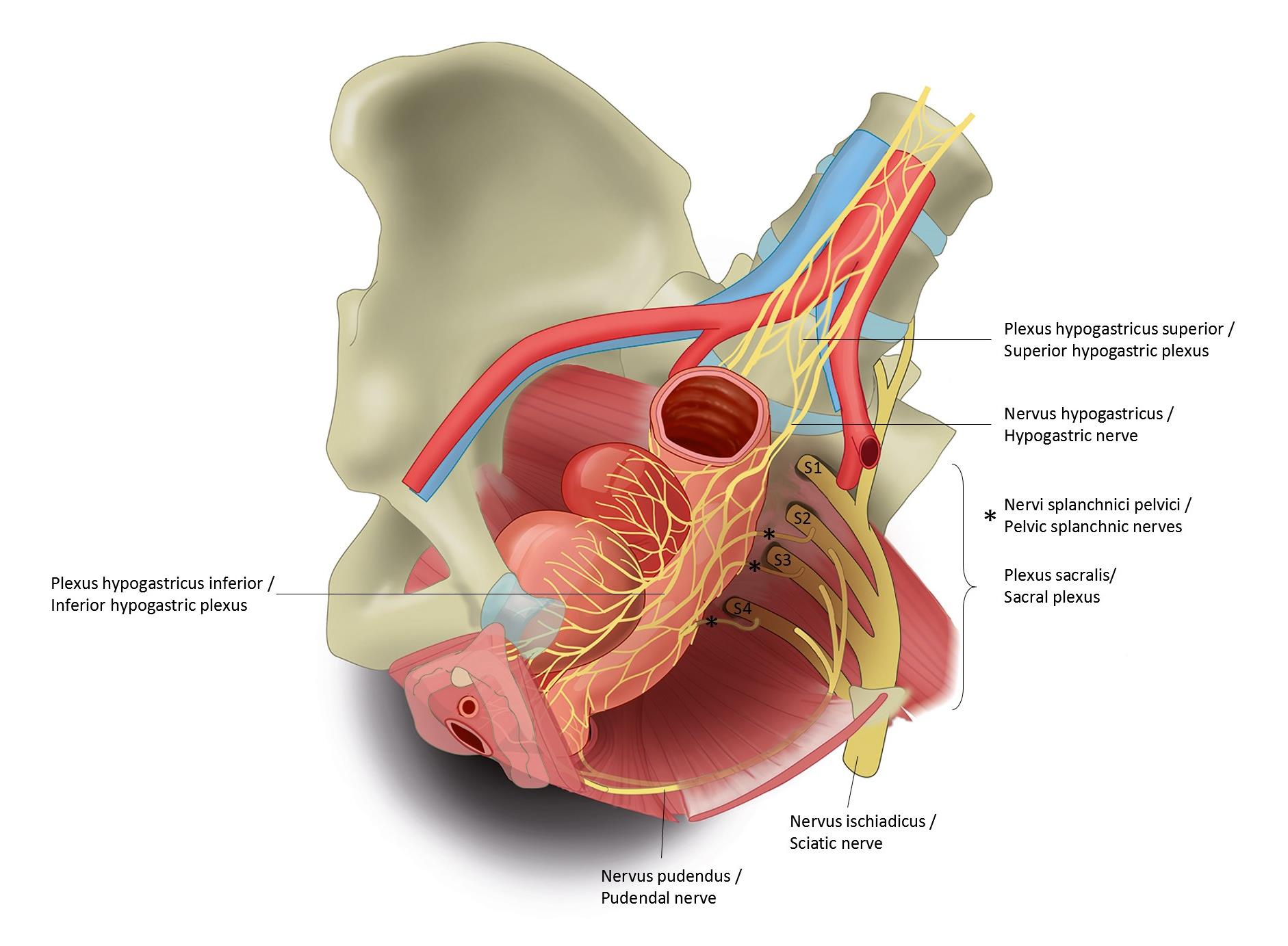
Anterior view of female pelvis; internal organs and innervation. From anterior to posterior: bladder, uterus, rectum. The aortic bifurcation, common iliac arteries and external and internal iliac arteries are shown. On the right the bony hemipelvis has been removed and the sacral plexus is shown, lying in front of the coccygeus muscle. The course of the pudendal nerve is depicted. On the lateral side of the pelvic organs the pelvic plexus can be seen (inferior hypogastric plexus). It receives nerve fibers from the sacral ventral rami and the superior hypogastric plexus, seen anterior to the aortic bifurcation. No labels.
ERRATUM: in reality, S4 does not pass into the sciatic nerve.
Illustration by Ron Slagter and Marco DeRuiter for course 'Surgical Anatomy of the lesser pelvis' by the 'Urologisch Opleidings Instituut', the Netherlands.
Slightly edited and labeled by Paul Gobée
ERRATUM: in reality, S4 does not pass into the sciatic nerve.
Illustration by Ron Slagter and Marco DeRuiter for course 'Surgical Anatomy of the lesser pelvis' by the 'Urologisch Opleidings Instituut', the Netherlands.
Slightly edited and labeled by Paul Gobée
Anatomical structures in item:
Uploaded by: admin
Netherlands, Leiden – Leiden University Medical Center, Leiden University
Plexus nervosus hypogastricus inferior
Plexus nervosus vesicalis
Nervus pudendus
Plexus nervosus hypogastricus superior
Nervi splanchnici pelvici
Musculus coccygeus
Musculus levator ani
Creator(s)/credit: Ron Slagter NZIMBI, medical illustrator, LUMC; Prof. Marco DeRuiter PhD, anatomist, LUMC; O. Paul Gobée MD, anatomist, LUMC
Requirements for usage
You are free to use this item if you follow the requirements of the license:  View license
 View license
 If you use this item you should credit it as follows:
- For usage in print - copy and paste the line below:
- For digital usage (e.g. in PowerPoint, Impress, Word, Writer) - copy and paste the line below (optionally add the license icon):
"Anterior view of female pelvis; internal organs and innervation - Latin and English labels" at AnatomyTOOL.org by Ron Slagter, LUMC, Marco DeRuiter, LUMC and O. Paul Gobée, LUMC, license: Creative Commons Attribution-NonCommercial-ShareAlike
"Anterior view of female pelvis; internal organs and innervation - Latin and English labels" by Ron Slagter, LUMC, Marco DeRuiter, LUMC and O. Paul Gobée, LUMC, license: CC BY-NC-SA


Comments