nid: 61473
Additional formats:
None available
Description:
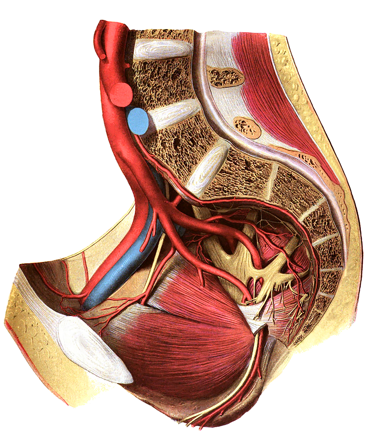
Blood vessels and nervus of the pelvic wall: lateral view. Labels and leader lines removed and colour brightened.
From 'Atlas and Textbook of Human Anatomy', 1909, Vol. 3, fig.568, by Johannes Sobotta and J. Playfair McMurrich. Artist: K. Hajek. Retrieved from Sobotta's Anatomy plates at Wikimedia. Possible original source: Sobotta's atlas at Hathitrust Digital library.
Image editing by dream_studio3.
From 'Atlas and Textbook of Human Anatomy', 1909, Vol. 3, fig.568, by Johannes Sobotta and J. Playfair McMurrich. Artist: K. Hajek. Retrieved from Sobotta's Anatomy plates at Wikimedia. Possible original source: Sobotta's atlas at Hathitrust Digital library.
Image editing by dream_studio3.
Anatomical structures in item:
Uploaded by: opgobee
Netherlands, Leiden – Leiden University Medical Center, Leiden University
Pelvis
Arteria mesenterica inferior
Bifurcatio aortae
Arteria iliaca communis
Vena iliaca communis
Arteria sacralis mediana
Arteria iliaca interna
Arteria iliolumbalis
Arteria iliaca externa
Arteria umbilicalis
Nervus obturatorius
Arteria obturatoria
Arteria cremasterica
Discus interpubicus
Arteria vesicalis inferior
Musculus obturatorius internus
Nervus pudendus
Nervus coccygeus
Plexus sacralis
Arteria glutea inferior
Arteria glutea superior
Truncus lumbosacralis
Arteriae sacrales laterales
Canalis pudendalis
Creator(s)/credit: Prof.dr. Johannes Sobotta, anatomist; dream_studio3 BA, image editing
Requirements for usage
You are free to use this item if you follow the requirements of the license:  View license
 View license
 If you use this item you should credit it as follows:
- For usage in print - copy and paste the line below:
- For digital usage (e.g. in PowerPoint, Impress, Word, Writer) - copy and paste the line below (optionally add the license icon):
"Sobotta 1909 fig.568 - Blood vessels and nervus of the pelvic wall - no labels" at AnatomyTOOL.org by Johannes Sobotta and dream_studio3, license: Creative Commons Attribution-ShareAlike


Comments