nid: 61139
Additional formats:
None available
Description:
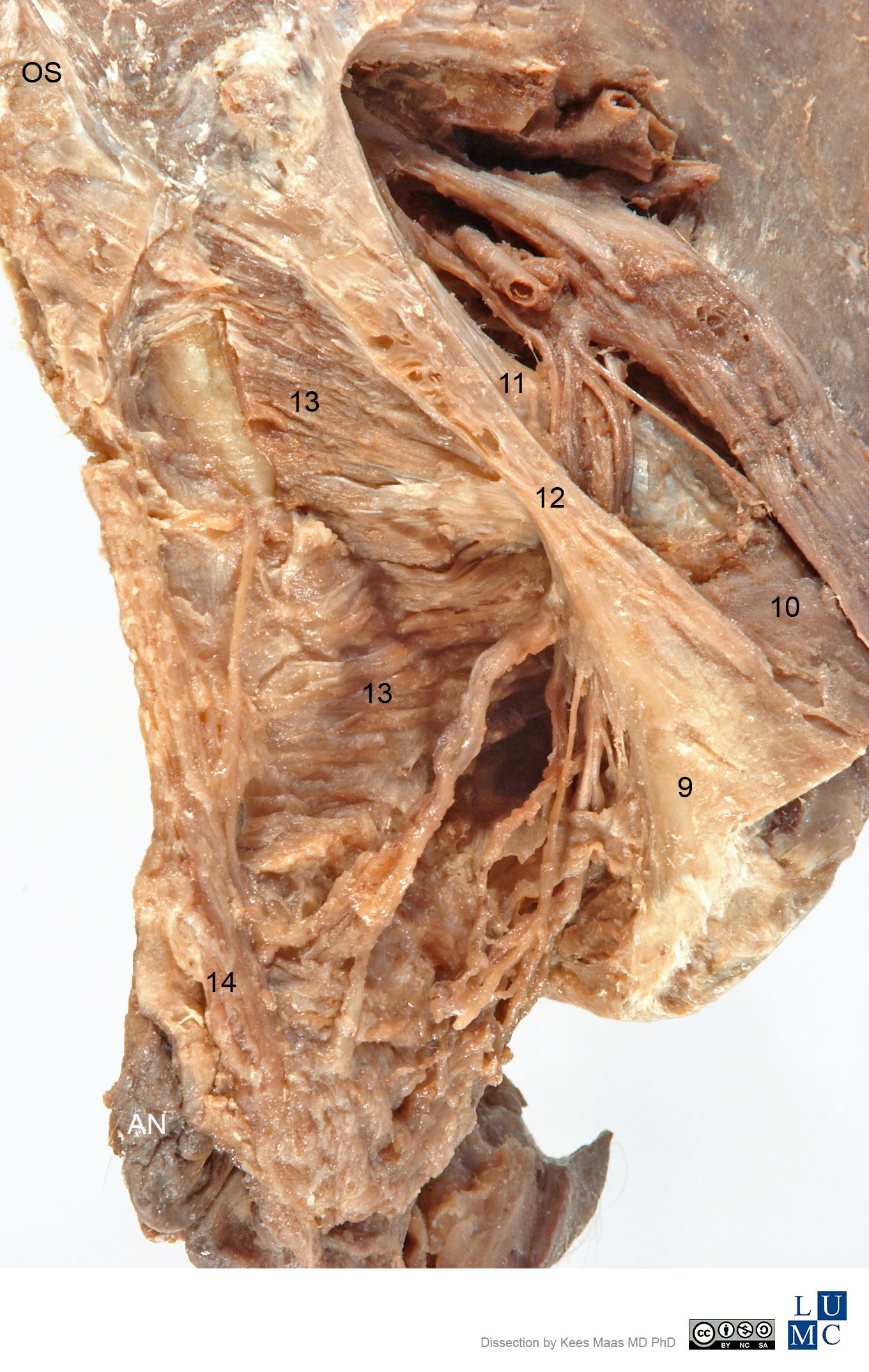
Posterior lateral view of the female pelvis. The specimen shows the pelvis from posterior lateral, especially large ligaments and muscles. Labels.
The key of the labels can be found in Leiden, Maas - Pelvis plastination specimens - English labels
The key of the labels can be found in Leiden, Maas - Pelvis plastination specimens - English labels
Anatomical structures in item:
Uploaded by: rva
Netherlands, Leiden – Leiden University Medical Center, Leiden University
Pelvis
Os sacrum [vertebrae sacrales I - V]
Anus
Tuber ischiadicum
Spina ischiadica
Ligamentum sacrospinale
Ligamentum sacrotuberale
Musculus levator ani
Musculus sphincter ani externus
Creator(s)/credit: Kees (C.P.) Maas MD, PhD, dissection, LUMC; Prof. Marco C. DeRuiter PhD, anatomist, professor of Clinical and Experimental Anatomy, LUMC; J. Lens, medical photographer
Requirements for usage
You are free to use this item if you follow the requirements of the license:  View license
 View license
 If you use this item you should credit it as follows:
- For usage in print - copy and paste the line below:
- For digital usage (e.g. in PowerPoint, Impress, Word, Writer) - copy and paste the line below (optionally add the license icon):
"Leiden, Maas Photo 18 - Posterior lateral view of the female pelvis (plastination specimen) - labels" at AnatomyTOOL.org by Kees (C.P.) Maas, LUMC, Marco C. DeRuiter, LUMC and J. Lens, license: Creative Commons Attribution-NonCommercial-ShareAlike
"Leiden, Maas Photo 18 - Posterior lateral view of the female pelvis (plastination specimen) - labels" by Kees (C.P.) Maas, LUMC, Marco C. DeRuiter, LUMC and J. Lens, license: CC BY-NC-SA


Comments