nid: 60568
Additional formats:
None available
Description:
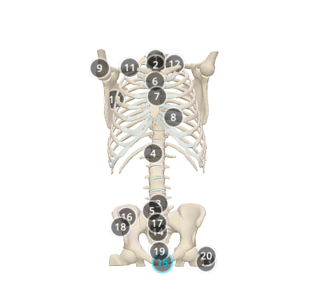
This model, depicts the bony structures of the thorax and abdomen. It was created in Pixologic Zbrush, based on CT data (from which subtools were extracted), specimen in the dissection room and references in anatomy books.
Anatomical structures in item:
Uploaded by: rva
Netherlands, Leiden – Leiden University Medical Center, Leiden University
Vertebra prominens [C VII]
Vertebrae thoracicae (TI-TXII)
Discus intervertebralis
Vertebrae lumbales (LI-LV)
Manubrium sterni
Sternum
Cartilago costalis
Humerus
Scapula
Clavicula
Costa prima [I]
Femur
Os sacrum [vertebrae sacrales I - V]
Symphysis pubica
Ilium
Promontorium ossis sacri
Spina iliaca anterior superior
Coccyx [vertebrae coccygeae I-IV]
Trochanter major
Creator(s)/credit: Anna Sieben MSc, medical illustrator
Requirements for usage
You are free to use this item if you follow the requirements of the license:  View license
 View license
 If you use this item you should credit it as follows:
- For usage in print - copy and paste the line below:
- For digital usage (e.g. in PowerPoint, Impress, Word, Writer) - copy and paste the line below (optionally add the license icon):
"Groningen - 3D model Thoracic & abdominal skeleton" at AnatomyTOOL.org by Anna Sieben, license: Creative Commons Attribution-NonCommercial-ShareAlike. Reviewed by: Cyril Luman, Dr. Walter Noordzij
"Groningen - 3D model Thoracic & abdominal skeleton" by Anna Sieben, license: CC BY-NC-SA. Reviewed by: Cyril Luman, Dr. Walter Noordzij


Comments