nid: 60424
Additional formats:
None available
Description:
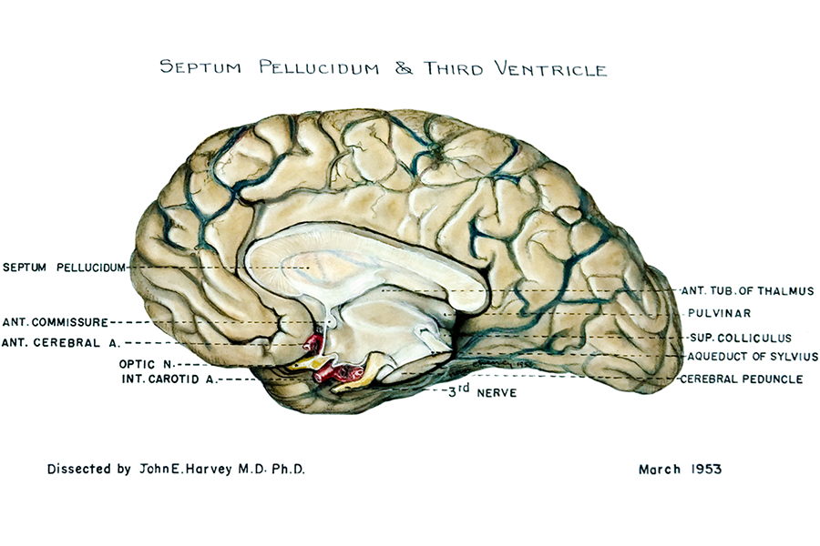
Third ventricle and septum pellucidum. The third ventricle of the brain can be seen, with its adjacent structures. English labels.
Retrieved from website Clinical Anatomy of the University of British Columbia.
Retrieved from website Clinical Anatomy of the University of British Columbia.
Anatomical structures in item:
Uploaded by: rva
Netherlands, Leiden – Leiden University Medical Center, Leiden University
Encephalon
Septum pellucidum
Commissura anterior
Arteria cerebri anterior
Arteria carotis interna
Tuberculum anterius thalami
Pulvinar thalami
Colliculus superior
Aqueductus mesencephali
Pedunculus cerebri
Creator(s)/credit: A.G.L. (Nan) Cheney, medical illustrator, UBC; Dr. John E. Harvey PhD, MD, UBC
Requirements for usage
You are free to use this item if you follow the requirements of the license:  View license
 View license
 If you use this item you should credit it as follows:
- For usage in print - copy and paste the line below:
- For digital usage (e.g. in PowerPoint, Impress, Word, Writer) - copy and paste the line below (optionally add the license icon):
"U.Br.Columbia - Drawing Third ventricle and septum pellucidum - English labels" at AnatomyTOOL.org by A.G.L. (Nan) Cheney, UBC and John E. Harvey, UBC, license: Creative Commons Attribution-NonCommercial-ShareAlike. Source: website Clinical Anatomy, http://www.clinicalanatomy.ca
"U.Br.Columbia - Drawing Third ventricle and septum pellucidum - English labels" by A.G.L. (Nan) Cheney, UBC and John E. Harvey, UBC, license: CC BY-NC-SA. Source: website Clinical Anatomy, http://www.clinicalanatomy.ca


Comments