
nid: 60394
Additional formats:
None available
Description:
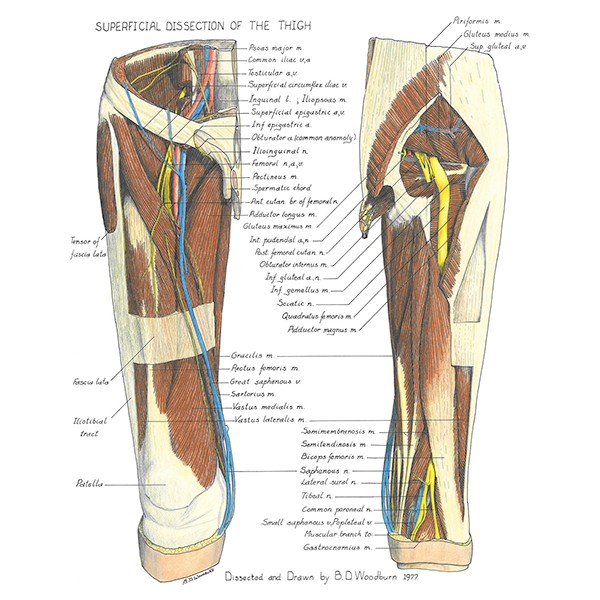
Superficial anatomy of the thigh. The anatomy of the thigh, especially the relation of the muscles with adjecent structures can be appreciated. English labels.
Retrieved from website Clinical Anatomy of the University of British Columbia.
Retrieved from website Clinical Anatomy of the University of British Columbia.
Anatomical structures in item:
Uploaded by: rva
Netherlands, Leiden – Leiden University Medical Center, Leiden University
Femur
Vena iliaca communis
Arteria iliaca communis
Vena circumflexa ilium superficialis
Arteria epigastrica superficialis
Vena epigastrica superficialis
Arteria epigastrica inferior
Arteria obturatoria
Nervus ilioinguinalis
Musculus pectineus
Rami cutanei anteriores nervus femoralis
Musculus adductor longus
Musculus tensor fasciae latae
Fascia lata
Tractus iliotibialis
Musculus vastus lateralis
Musculus vastus medialis
Musculus sartorius
Vena saphena magna
Musculus rectus femoris
Musculus gracilis
Musculus adductor magnus
Musculus quadratus femoris
Musculus gemellus inferior
Arteria glutea inferior
Nervus gluteus inferior
Musculus obturatorius internus
Nervus cutaneus femoris posterior
Arteria pudenda interna
Musculus gluteus maximus
Musculus gluteus medius
Venae gluteae superiores
Arteria glutea superior
Requirements for usage
You are free to use this item if you follow the requirements of the license:  View license
View license If you use this item you should credit it as follows:
- For usage in print - copy and paste the line below:
- For digital usage (e.g. in PowerPoint, Impress, Word, Writer) - copy and paste the line below (optionally add the license icon):
"U.Br.Columbia - Drawing Superficial anatomy of the thigh - English labels" at AnatomyTOOL.org by , license: Creative Commons Attribution-NonCommercial-ShareAlike. Created for: Department of Anatomy (now Department of Cellular and Physiological Sciences) at the University of British Columbia. Source: website Clinical Anatomy, http://www.clinicalanatomy.ca
"U.Br.Columbia - Drawing Superficial anatomy of the thigh - English labels" by , license: CC BY-NC-SA. Created for: Department of Anatomy (now Department of Cellular and Physiological Sciences) at the University of British Columbia. Source: website Clinical Anatomy, http://www.clinicalanatomy.ca


Comments