
nid: 60369
Additional formats:
None available
Description:
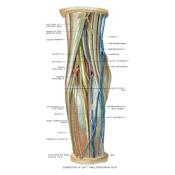
Posterior view of the knee, popliteal fossa. The structures of the popliteal fossa can be seen in this figure. English labels.
Retrieved from website Clinical Anatomy of the University of British Columbia.
Retrieved from website Clinical Anatomy of the University of British Columbia.
Anatomical structures in item:
Uploaded by: rva
Netherlands, Leiden – Leiden University Medical Center, Leiden University
Genu
Musculus biceps femoris
Fossa poplitea
Nervus ischiadicus
Arteria poplitea
Nervus tibialis
Musculus gastrocnemius
Nervus suralis
Ramus communicans nervus peronei communis cum nervus cutaneo surae mediali
Musculus soleus
Musculus fibularis longus
Vena saphena parva
Musculus gastrocnemius
Articulatio genus
Vena saphena magna
Vena saphena parva
Musculus sartorius
Musculus semitendinosus
Musculus semimembranosus
Musculus gracilis
Creator(s)/credit: A.G.L. (Nan) Cheney, medical illustrator, UBC; G.E. Singer M.D.C.M. M.Sc., UBC
Requirements for usage
You are free to use this item if you follow the requirements of the license:  View license
View license If you use this item you should credit it as follows:
- For usage in print - copy and paste the line below:
- For digital usage (e.g. in PowerPoint, Impress, Word, Writer) - copy and paste the line below (optionally add the license icon):
"U.Br.Columbia - Drawing Posterior view of the knee, popliteal fossa - English labels" at AnatomyTOOL.org by A.G.L. (Nan) Cheney, UBC and G.E. Singer, UBC, license: Creative Commons Attribution-NonCommercial-ShareAlike. Source: website Clinical Anatomy, http://www.clinicalanatomy.ca
"U.Br.Columbia - Drawing Posterior view of the knee, popliteal fossa - English labels" by A.G.L. (Nan) Cheney, UBC and G.E. Singer, UBC, license: CC BY-NC-SA. Source: website Clinical Anatomy, http://www.clinicalanatomy.ca


Comments