
nid: 60338
Additional formats:
None available
Description:
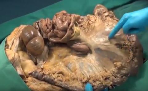
The detachment of peritoneal adherences as forms the basis of many gastrointestinal surgical procedures is shown in a dissection specimen. The greater omentum is detached from the transverse colon, giving access to the omental bursa. The descending colon and mesocolon are detached, the so-called left visceral rotation. The Cattel-Braasch manoeuvre - detaching and lifting the ascending (meso) colon and small intestines, to give access to the peritoneal back wall - is demonstrated. This also unveils the hidden part of the duodenum. Finally, it is shown how the mesocolons and the mesentery of the small intestine form a continuity that rotated with the intestines embryologically.
This video is from the MOOC 'Anatomy of the abdomen and pelvis: a journey from basis to clinic' by the Leiden University Medical Center.
This video is also availabale at https://youtu.be/BKMaeT1VIho
This video is from the MOOC 'Anatomy of the abdomen and pelvis: a journey from basis to clinic' by the Leiden University Medical Center.
This video is also availabale at https://youtu.be/BKMaeT1VIho
Anatomical structures in item:
Uploaded by: rjjvisser
Netherlands, Leiden – Leiden University Medical Center, Leiden University
Ventriculus
Omentum majus
Colon
Ileum
Jejunum
Peritoneum
Mesocolon ascendens
Mesocolon descendens
Mesocolon
Mesocolon transversum
Radix mesenterii
Mesentery
Duodenum
Peritoneum parietale
Creator(s)/credit: O. Paul Gobée MD, Anatomist, LUMC; J. de Roeck-den Boeft, video, LUMC
Requirements for usage
You are free to use this item if you follow the requirements of the license:  View license
View license If you use this item you should credit it as follows:
- For usage in print - copy and paste the line below:
- For digital usage (e.g. in PowerPoint, Impress, Word, Writer) - copy and paste the line below (optionally add the license icon):
"Leiden MOOC 4.13 - Video Anatomy on the table: peritoneal structures" at AnatomyTOOL.org by O. Paul Gobée, LUMC and J. de Roeck-den Boeft, LUMC, license: Creative Commons Attribution-NonCommercial-ShareAlike
"Leiden MOOC 4.13 - Video Anatomy on the table: peritoneal structures" by O. Paul Gobée, LUMC and J. de Roeck-den Boeft, LUMC, license: CC BY-NC-SA


Comments