
nid: 60126
Additional formats:
None available
Description:
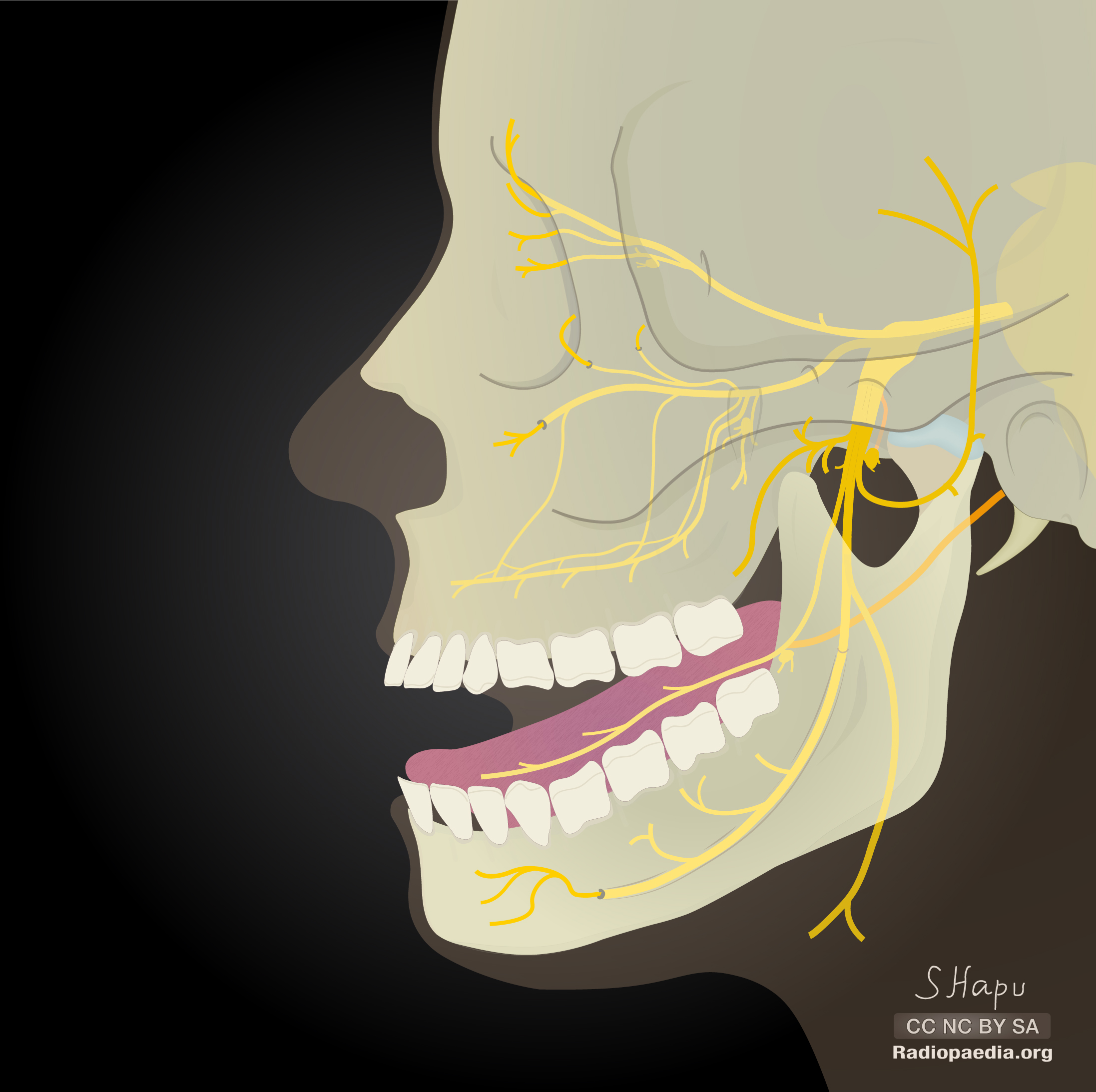
Main branches of the trigeminal nerve. This image shows the main branches (motor and sensor) of the trigeminal nerve (CN V). No labels.
Case courtesy of Dr Sachintha Hapugoda, Radiopaedia.org. From the case rID: 54722
Case courtesy of Dr Sachintha Hapugoda, Radiopaedia.org. From the case rID: 54722
Anatomical structures in item:
Uploaded by: rva
Netherlands, Leiden – Leiden University Medical Center, Leiden University
Nervus frontalis
Nervus lacrimalis
Nervus nasociliaris
Ganglion ciliare
Fissura orbitalis superior
Nervus trigeminus [V]
Nervus ophthalmicus [Va]
Nervus maxillaris [Vb]
Foramen rotundum
Nervus zygomaticus
Nervus infraorbitalis
Fossa pterygopalatina
Foramen infraorbitale
Ganglion pterygopalatinum
Nervi alveolares superiores
Nervus mandibularis [Vc]
Foramen ovale
Ganglion oticum
Nervi temporales profundi
Nervus pterygoideus lateralis
Nervus massetericus
Nervus auriculotemporalis
Nervus buccalis
Ganglion submandibulare
Nervus lingualis
Canalis mandibulae
Nervus alveolaris inferior
Nervus mentalis
Foramen mentale
Nervus mylohyoideus
Creator(s)/credit: Dr Sachintha Hapugoda MB.BS, MMed
Requirements for usage
You are free to use this item if you follow the requirements of the license:  View license
View license If you use this item you should credit it as follows:
- For usage in print - copy and paste the line below:
- For digital usage (e.g. in PowerPoint, Impress, Word, Writer) - copy and paste the line below (optionally add the license icon):
"Radiopaedia - Drawing Main branches of the trigeminal nerve - no labels" at AnatomyTOOL.org by Sachintha Hapugoda, license: Creative Commons Attribution-NonCommercial-ShareAlike


Comments