nid: 59994
Additional formats:
None available
Description:
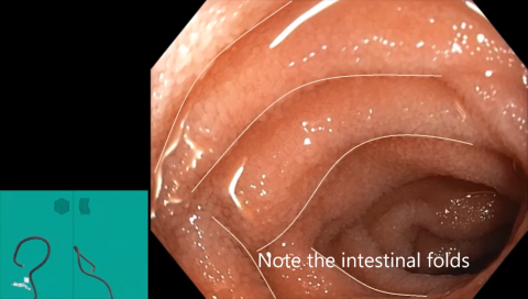
This video shows a part of a colonoscopy, with normal anatomy: it starts in the terminal ileum, where the circular folds and the intestinal villi can be seen and are clearly indicated. Then the scope is gradually retracted, passing the ileocaecal junction to arrive in the caecum. In the ascending colon the semilunar folds can be seen and are indicated, one can also see the absence of villi here. The clear endoscope imagery with high magnification in a life person, allows to see the individual villi and the difference between the circular and the semilunar folds in a way that is impossible to attain in an anatomical dissection lab. The video is created by A.M.J. Langers, MD, PhD, gastroenterologist, dept. of Gastroenterology, and edited by A.L. Schoenmakers, MD, dept. of Anatomy and Embryology, both at Leiden University Medical Center, the Netherlands.
Anatomical structures in item:
Uploaded by: opgobee
Netherlands, Leiden – Leiden University Medical Center, Leiden University
Ileum
Pars terminalis
Caecum
Villi intestinales intestini tenuis
Plicae circulares intestini tenuis
Plicae semilunares coli
Ileocecal junction
Creator(s)/credit: Alexandra M.J. Langers MD, PhD, Gastroenterologist, LUMC; Alieke L. Schoenmakers MD, resident, video editing, LUMC
Requirements for usage
You are free to use this item if you follow the requirements of the license:  View license
 View license
 If you use this item you should credit it as follows:
- For usage in print - copy and paste the line below:
- For digital usage (e.g. in PowerPoint, Impress, Word, Writer) - copy and paste the line below (optionally add the license icon):
"Leiden - Video Colonoscopy: transition from ileum to caecum - English labels" at AnatomyTOOL.org by Alexandra M.J. Langers, LUMC and Alieke L. Schoenmakers, LUMC, license: Creative Commons Attribution-NonCommercial-ShareAlike


Comments