
nid: 59901
Additional formats:
None available
Description:
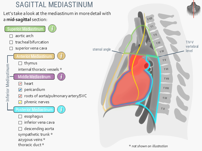
The middle mediastinum.This image shows a sagittal view of the mediastinum. The middle mediastinum is part of the inferior mediastinum together with the anterior and posterior mediastinum. The superior, anterior, middle and posterior mediastinum are marked with respectively a green, orange, purple and light blue line. The middle mediastinum contains the heart (in light red), the pericardium (blue), roots of aorta, pulmonary artery and superior vena cava (red) and phrenic nerves (yellow). English labels. Retrieved from the interactive module Around the heart: Mediastinum from the website Clinical Anatomy of the University of British Columbia.
Anatomical structures in item:
Uploaded by: rva
Netherlands, Leiden – Leiden University Medical Center, Leiden University
Cor
Mediastinum
Mediastinum inferius
Mediastinum medium
Cavitas thoracis
Cor
Pericardium
Bulbus aortae
Nervus phrenicus
Creator(s)/credit: Prof. Claudia Krebs MD, PhD, anatomist, UBC; Monika Fejtek, digital media technologist, UBC; Rebecca Comeau MD, UBC; Dr. Olusegun Oyedele; Dr. Paul Rea; Daniel McClusky; Iskander Afiq Mohamad Hashim; Jenna Woods
Requirements for usage
You are free to use this item if you follow the requirements of the license:  View license
View license If you use this item you should credit it as follows:
- For usage in print - copy and paste the line below:
- For digital usage (e.g. in PowerPoint, Impress, Word, Writer) - copy and paste the line below (optionally add the license icon):
"U.Br.Columbia - Drawing The middle mediastinum - English labels" at AnatomyTOOL.org by Claudia Krebs, UBC, Monika Fejtek, UBC, Rebecca Comeau, UBC et al, license: Creative Commons Attribution-NonCommercial-ShareAlike. Source: website Clinical Anatomy, http://www.clinicalanatomy.ca
"U.Br.Columbia - Drawing The middle mediastinum - English labels" by Claudia Krebs, UBC, Monika Fejtek, UBC, Rebecca Comeau, UBC et al, license: CC BY-NC-SA. Source: website Clinical Anatomy, http://www.clinicalanatomy.ca


Comments