nid: 59850
Additional formats:
None available
Description:
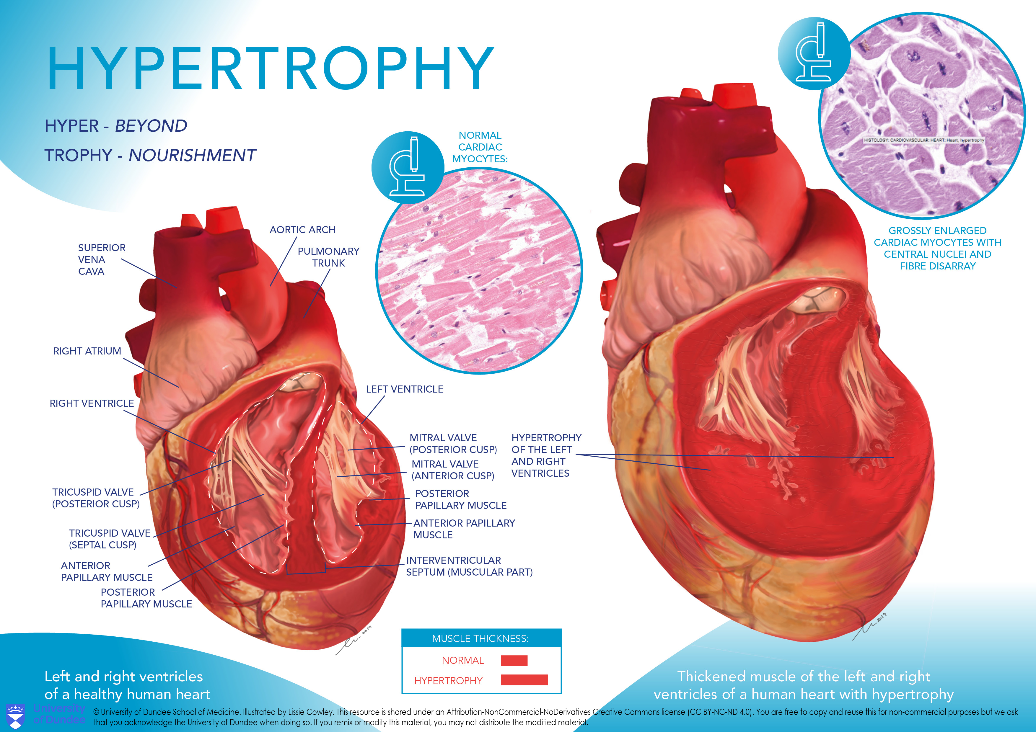
Hypertrophy of the heart. On the left, a healthy human heart is shown, whereas on the right, an enlarged heart is shown. English labels. NOTE: THIS IMAGE IS UNDER A NON-DERIVATIVE LICENSE. THIS MEANS THAT IF YOU REMIX OR REVISE THIS MATERIAL YOU MAY NOT DISTRIBUTE THE MODIFIED MATERIAL.
Anatomical structures in item:
Uploaded by: rva
Netherlands, Leiden – Leiden University Medical Center, Leiden University
Cor
Vena cava superior
Atrium dextrum
Ventriculus dexter
Valva tricuspidalis
Cuspis posterior valvae atrioventricularis dextrae
Cuspis septalis valvae atrioventricularis dextrae
Musculus papillaris anterior ventriculi dextri
Musculus papillaris posterior ventriculi sinistri
Musculus papillaris
Musculi papillares cordis
Septum interventriculare
Musculus papillaris anterior ventriculi sinistri
Musculus papillaris posterior ventriculi dextri
Valva mitralis
Cuspis posterior valvae atrioventricularis sinistri
Cuspis anterior valvae atrioventricularis sinistri
Ventriculus sinister
Truncus pulmonalis
Arcus aortae
Creator(s)/credit: Lissie Cowley MSc, medical illustrator
Requirements for usage
You are free to use this item if you follow the requirements of the license:  View license
 View license
 If you use this item you should credit it as follows:
- For usage in print - copy and paste the line below:
- For digital usage (e.g. in PowerPoint, Impress, Word, Writer) - copy and paste the line below (optionally add the license icon):
"Dundee - Drawing Hypertrophy of the heart - English labels" at AnatomyTOOL.org by Lissie Cowley, © University of Dundee School of Medicine, license: Creative Commons Attribution-NonCommercial-NoDerivs
"Dundee - Drawing Hypertrophy of the heart - English labels" by Lissie Cowley, © University of Dundee School of Medicine, license: CC BY-NC-ND


Comments