
nid: 59799
Additional formats:
None available
Description:
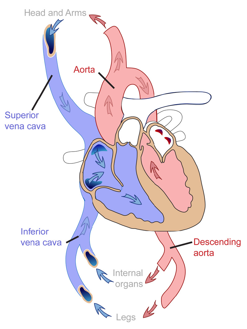
The systemic circulation. Systemic circulation is the part of the circulatory system that carries blood between the heart and body. English labels.
Anatomical structures in item:
Uploaded by: rva
Netherlands, Leiden – Leiden University Medical Center, Leiden University
Aorta
Cor
Vena cava superior
Vena cava inferior
Aorta descendens
Creator(s)/credit: Mariana Ruiz Villarreal (Wikimedia: LadyofHats)
Requirements for usage
You are free to use this item if you follow the requirements of the license:  View license
View license If you use this item you should credit it as follows:
- For usage in print - copy and paste the line below:
- For digital usage (e.g. in PowerPoint, Impress, Word, Writer) - copy and paste the line below (optionally add the license icon):
"Human Biology fig. 1.58 - The systemic circulation - English labels" at AnatomyTOOL.org by Mariana Ruiz Villarreal (Wikimedia: LadyofHats), license: Creative Commons Attribution-NonCommercial. Source: book ‘Human Biology’, https://textbookequity.org/Textbooks/HumanBiologyCK12.pdf.
"Human Biology fig. 1.58 - The systemic circulation - English labels" by Mariana Ruiz Villarreal (Wikimedia: LadyofHats), license: CC BY-NC. Source: book ‘Human Biology’, https://textbookequity.org/Textbooks/HumanBiologyCK12.pdf.


Comments