
nid: 59717
Additional formats:
None available
Description:
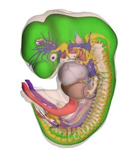
A series of 14 interactive 3D pdf files with detailed 3D models of human embryos and their organ systems in Carnegie stages 7 to 23 (15-60 days of development). The models are reconstructions based on segmentation of 15,000 manually annotated sections of 34 embryo's of the renowned Carnegie embryo collection. To view the 3D models, download the files and view them in Acrobat Reader. Individual structures in the embryos can be shown and hidden. The atlas was created by students and embryologists, led by Bernadette S. de Bakker, MD, PhD. of the Department of Anatomy, Embryology & Physiology of the Academic Medical Center (AMC) in Amsterdam, the Netherlands.
Anatomical structures in item:
Uploaded by: opgobee
Netherlands, Leiden – Leiden University Medical Center, Leiden University
Cor
Pulmones
Encephalon
Abdomen
Creator(s)/credit: Bernadette S. de Bakker MD, PhD, anatomist, AMC; 3D embryo atlas team, AMC
Requirements for usage
You are free to use this item if you follow the requirements of the license:  View license
View license If you use this item you should credit it as follows:
- For usage in print - copy and paste the line below:
- For digital usage (e.g. in PowerPoint, Impress, Word, Writer) - copy and paste the line below (optionally add the license icon):
"Amsterdam - 3D Atlas of Human Embryology" at AnatomyTOOL.org by Bernadette S. de Bakker, AMC and 3D embryo atlas team, AMC, license: Creative Commons Attribution-NonCommercial-NoDerivs
"Amsterdam - 3D Atlas of Human Embryology" by Bernadette S. de Bakker, AMC and 3D embryo atlas team, AMC, license: CC BY-NC-ND


Comments