
nid: 59623
Additional formats:
None available
Description:
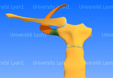
In this video the anatomy of the sternoclavicular (SC) joint is shown and explained. This joint connects the sternum, the medial part of the clavicule (and the first costa). This video is in the "3D Anatomy Lyon" series from the Université Claude Bernard in Lyon, France. NOTE: THIS VIDEO IS UNDER A NON-DERIVATIVE LICENSE. THIS MEANS THAT IF YOU REMIX, TRANSFORM, OR BUILD UPON THE MATERIAL, YOU MAY NOT DISTRIBUTE THE MODIFIED MATERIAL.
Anatomical structures in item:
Uploaded by: rva
Netherlands, Leiden – Leiden University Medical Center, Leiden University
Articulatio sternoclavicularis
Manubrium sterni
Sternum
Clavicula
Facies articularis sternalis claviculae
Extremitas sternalis claviculae
Costa prima [I]
Ligamentum sternoclaviculare anterius
Ligamentum sternoclaviculare posterius
Ligamentum interclaviculare
Ligamentum costoclaviculare
Musculus subclavius
Creator(s)/credit: Patrice Thiriet PhD, anatomist, project leader of Anatomie 3D Lyon; Jean-Michel Grand; Olivier Rastello, multimedia expert; Nora van Reeth, project manager; Christophe Batier, technical director
Requirements for usage
You are free to use this item if you follow the requirements of the license:  View license
View license If you use this item you should credit it as follows:
- For usage in print - copy and paste the line below:
- For digital usage (e.g. in PowerPoint, Impress, Word, Writer) - copy and paste the line below (optionally add the license icon):
"3D Anatomy Lyon: The sternoclavicular joint - video of 3D model" at AnatomyTOOL.org by Patrice Thiriet, Jean-Michel Grand, Olivier Rastello et al, license: Creative Commons Attribution-NonCommercial-NoDerivs. video in the "Anatomie 3D Lyon" series at https://www.youtube.com/channel/UC9LucUID-BUjL_c8oAT3vHQ/
"3D Anatomy Lyon: The sternoclavicular joint - video of 3D model" by Patrice Thiriet, Jean-Michel Grand, Olivier Rastello et al, license: CC BY-NC-ND. video in the "Anatomie 3D Lyon" series at https://www.youtube.com/channel/UC9LucUID-BUjL_c8oAT3vHQ/


Comments