nid: 59569
Additional formats:
None available
Description:
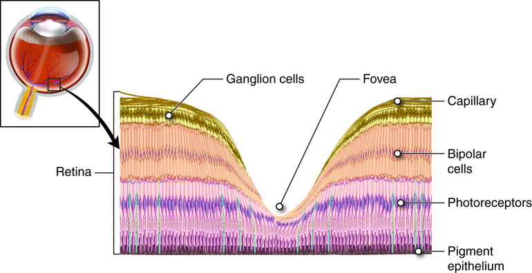
Anatomy of the fovea. At the exact center of the retina is a point where light is focused by the lens and the greatest visual acuity is found. This is known as the fovea and it is a small dimple in the layers of the retina where there are no blood vessels, ganglion cells or bipolar cells to interrupt light reaching the receptor cells. Because more light passes to the receptor cells at the fovea, it is in this region that visual acuity is the greatest. English labels.
Anatomical structures in item:
Uploaded by: rva
Netherlands, Leiden – Leiden University Medical Center, Leiden University
Retina
Stratum segmentorum externorum et internorum retinae
Stratum pigmentosum retinae
Fovea centralis
Creator(s)/credit: Cenveo
Requirements for usage
You are free to use this item if you follow the requirements of the license:  View license
 View license
 If you use this item you should credit it as follows:
- For usage in print - copy and paste the line below:
- For digital usage (e.g. in PowerPoint, Impress, Word, Writer) - copy and paste the line below (optionally add the license icon):
"Cenveo - Drawing Anatomy of the fovea - English labels" at AnatomyTOOL.org by Cenveo, license: Creative Commons Attribution
"Cenveo - Drawing Anatomy of the fovea - English labels" by Cenveo, license: CC BY


Comments