
nid: 59566
Additional formats:
None available
Description:
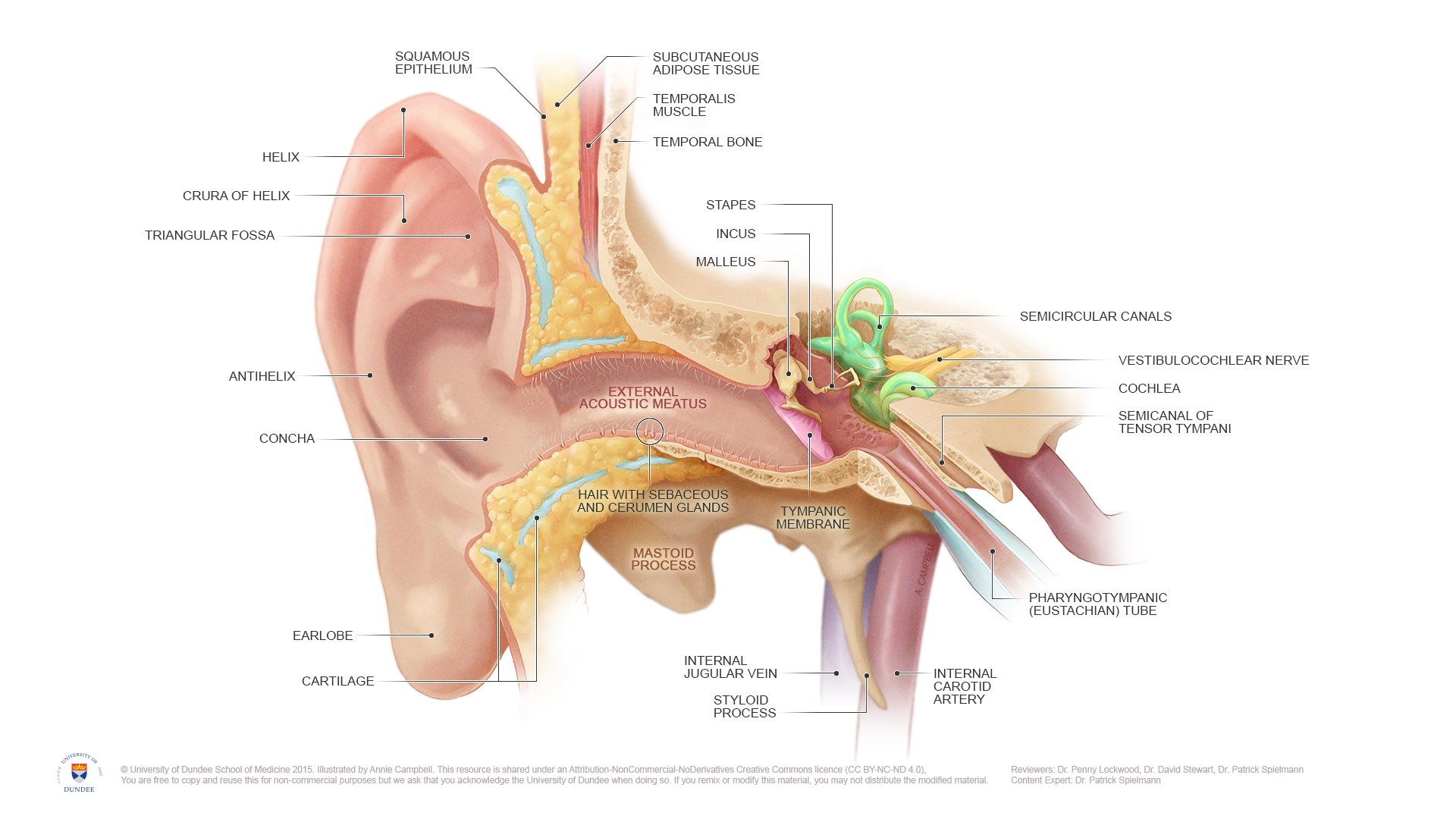
Anatomy of the ear. The anatomy of the inner and outer ear can be appreciated. English labels.
NOTE: THIS IMAGE IS UNDER A NON-DERIVATIVE LICENSE. THIS MEANS THAT IF YOU REMIX OR REVISE THIS MATERIAL YOU MAY NOT DISTRIBUTE THE MODIFIED MATERIAL.
NOTE: THIS IMAGE IS UNDER A NON-DERIVATIVE LICENSE. THIS MEANS THAT IF YOU REMIX OR REVISE THIS MATERIAL YOU MAY NOT DISTRIBUTE THE MODIFIED MATERIAL.
Anatomical structures in item:
Uploaded by: rva
Netherlands, Leiden – Leiden University Medical Center, Leiden University
Auris
Auris interna
Auris externa
Cochlea
Membrana tympanica
Helix
Crura antihelicis
Antihelix
Triangular fossa of antihelix
Concha auriculae
Lobulus auriculae
Meatus acusticus externus
Os temporale
Stapes
Incus
Malleus
Canales semicirculares
Nervus vestibulocochlearis [VIII]
Semicanalis musculi tensoris tympani
Tuba auditoria (auditiva)
Arteria carotis interna
Processus styloideus
Membrana tympanica
Creator(s)/credit: Annie Campbell MSc, medical illustrator
Requirements for usage
You are free to use this item if you follow the requirements of the license:  View license
View license If you use this item you should credit it as follows:
- For usage in print - copy and paste the line below:
- For digital usage (e.g. in PowerPoint, Impress, Word, Writer) - copy and paste the line below (optionally add the license icon):
"Dundee - Drawing Anatomy of the ear - English labels" at AnatomyTOOL.org by Annie Campbell, © University of Dundee School of Medicine, license: Creative Commons Attribution-NonCommercial-NoDerivs. Reviewed by: Dr. Penny Lockwood, Dr. David Stewart, Dr. Patrick Spielmann
"Dundee - Drawing Anatomy of the ear - English labels" by Annie Campbell, © University of Dundee School of Medicine, license: CC BY-NC-ND. Reviewed by: Dr. Penny Lockwood, Dr. David Stewart, Dr. Patrick Spielmann


Comments