
nid: 59275
Additional formats:
None available
Description:
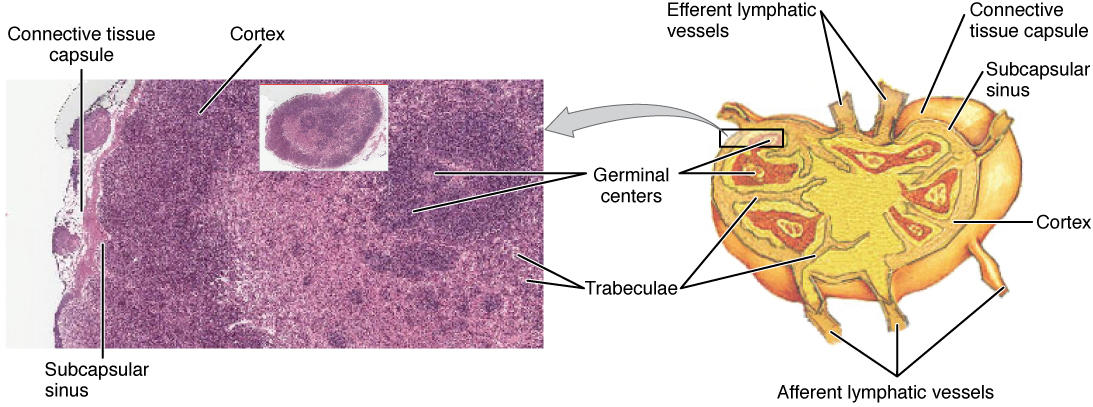
Structure and Histology of a Lymph Node. Lymph nodes are masses of lymphatic tissue located along the larger lymph vessels. The micrograph of the lymph nodes shows a germinal center, which consists of rapidly dividing B cells surrounded by a layer of T cells and other accessory cells. LM × 128. (Micrograph provided by the Regents of the University of Michigan Medical School © 2012). English labels. From OpenStax book 'Anatomy and Physiology', fig. 21.8.
Anatomical structures in item:
Uploaded by: Jorn IJkhout
Netherlands, Leiden – Leiden University Medical Center, Leiden University
Lymphonodus
Cortex nodi lymphoidei
Capsula nodi lymphoidei
Vas lymphaticum
Creator(s)/credit: OpenStax; Regents of U-M Medical School, UMich MedSchool
Requirements for usage
You are free to use this item if you follow the requirements of the license:  View license
View license If you use this item you should credit it as follows:
- For usage in print - copy and paste the line below:
- For digital usage (e.g. in PowerPoint, Impress, Word, Writer) - copy and paste the line below (optionally add the license icon):
"OpenStax AnatPhys fig.21.8 - Structure and Histology of a Lymph Node - English labels" at AnatomyTOOL.org by OpenStax and Regents of U-M Medical School, UMich MedSchool, license: Creative Commons Attribution. Source: book 'Anatomy and Physiology', https://openstax.org/details/books/anatomy-and-physiology.
"OpenStax AnatPhys fig.21.8 - Structure and Histology of a Lymph Node - English labels" by OpenStax and Regents of U-M Medical School, UMich MedSchool, license: CC BY. Source: book 'Anatomy and Physiology', https://openstax.org/details/books/anatomy-and-physiology.


Comments