
nid: 59194
Additional formats:
None available
Description:
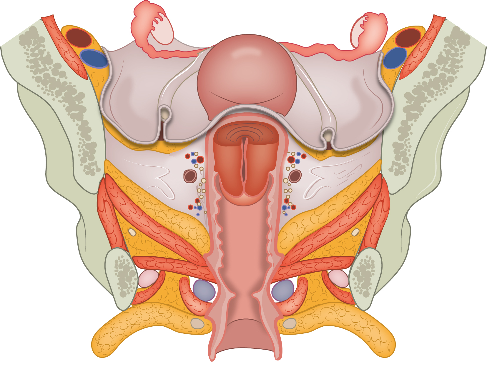
Coronal section of the female pelvis showing the cervix, vagina, cardinal ligament and levator ani muscle. No labels.
Illustration by Ron Slagter and Marco DeRuiter for course 'Surgical Anatomy of the lesser pelvis' by the 'Urologisch Opleidings Instituut', the Netherlands.
Illustration by Ron Slagter and Marco DeRuiter for course 'Surgical Anatomy of the lesser pelvis' by the 'Urologisch Opleidings Instituut', the Netherlands.
Anatomical structures in item:
Uploaded by: Siem Zethof
Netherlands, Leiden – Leiden University Medical Center, Leiden University
Ligamentum cardinale
Cervix uteri
Vagina
Musculus obturatorius internus
Musculus levator ani
Musculus transversus perinei profundus
Musculus ischiocavernosus
Musculus bulbospongiosus
Pelvis
Creator(s)/credit: Ron Slagter NZIMBI, medical illustrator, LUMC; Prof. Marco DeRuiter PhD, anatomist, LUMC
Requirements for usage
You are free to use this item if you follow the requirements of the license:  View license
View license If you use this item you should credit it as follows:
- For usage in print - copy and paste the line below:
- For digital usage (e.g. in PowerPoint, Impress, Word, Writer) - copy and paste the line below (optionally add the license icon):
"Coronal section of the female pelvis showing the cervix, vagina, cardinal ligament and levator ani muscle – no labels" at AnatomyTOOL.org by Ron Slagter, LUMC and Marco DeRuiter, LUMC, license: Creative Commons Attribution-NonCommercial-ShareAlike


Comments