
nid: 59186
Additional formats:
None available
Description:
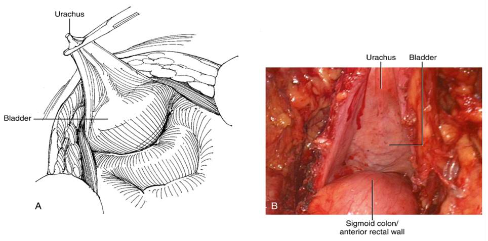
On the left; a V-shaped dissection of peritoneum superior to the bladder showing the median umbilical ligament, the urachus. On the right; a superior view of pelvic peritoneal cavity showing the urachus, bladder and anterior rectal wall. English labels. Smith, Howard, Preminger, Hinman's atlas of urologic surgery third ed 2012
Anatomical structures in item:
Uploaded by: Siem Zethof
Netherlands, Leiden – Leiden University Medical Center, Leiden University
Ligamentum umbilicale medianum
Vesica urinaria
Creator(s)/credit: Ron Slagter NZIMBI, medical illustrator, LUMC; Prof. Marco DeRuiter PhD, anatomist, LUMC
Requirements for usage
This item's license is unknown. Assume it is under copyright. You should request its owner permission to use it. However, provided the item is published online legally, you are free to link to it.

Comments