
nid: 59179
Additional formats:
None available
Description:
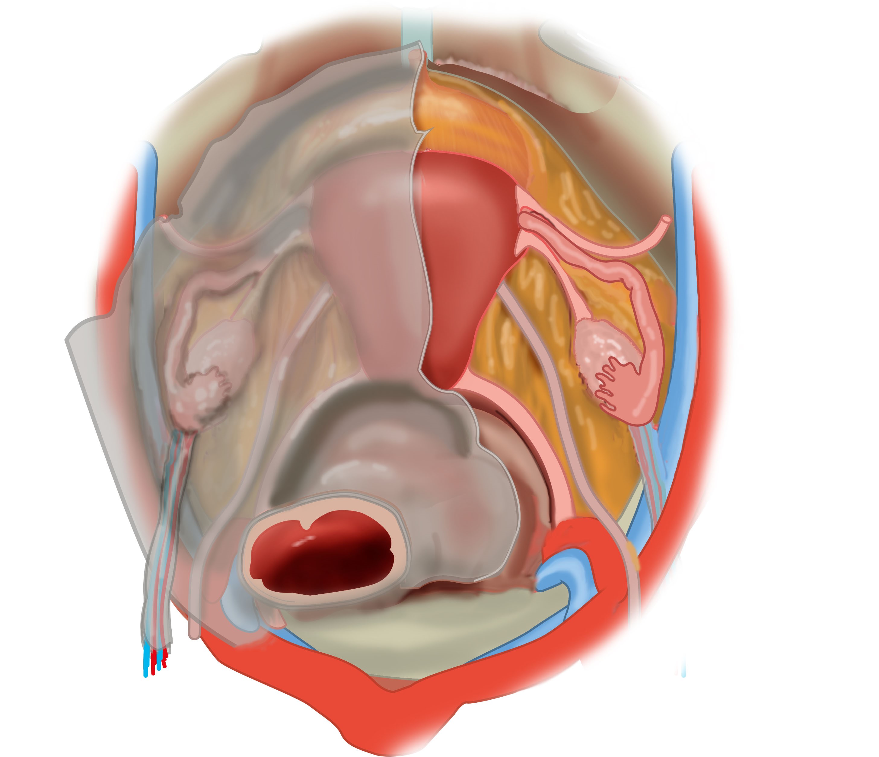
Superior view of female pelvis. Left; Peritoneum covering the uterus, it's ligaments and the ovaries is the broad ligament of the uterus. The recto-uterine pouch (Douglas) is depicted, extending laterally and posteriorly to form a pararectal fossa on each side of the rectum. Between the uterus and the bladder, a vesico-uterine pouch is formed. Right: Structures shown on the lateral side of the uterus, from anterior to posterior: round ligament of the uterus, uterine tube, ovarian ligament, ureter, uterosacral ligament. The ovary is connected to the lateral pelvic wall with the suspensory ligament of the ovary. The urachus lies anterosuperior on the bladder. No labels.
Illustration by Ron Slagter and Marco DeRuiter for course 'Surgical Anatomy of the lesser pelvis' by the 'Urologisch Opleidings Instituut', the Netherlands.
Illustration by Ron Slagter and Marco DeRuiter for course 'Surgical Anatomy of the lesser pelvis' by the 'Urologisch Opleidings Instituut', the Netherlands.
Anatomical structures in item:
Uploaded by: Siem Zethof
Netherlands, Leiden – Leiden University Medical Center, Leiden University
Ligamentum latum uteri
Excavatio vesicouterina
Excavatio rectouterina
Ligamentum teres uteri
Uterus
Tuba uterina (Salpinx)
Ligamentum ovarii proprium
Ureter
Ligamentum suspensorium ovarii
Creator(s)/credit: Ron Slagter NZIMBI, medical illustrator, LUMC; Prof. Marco DeRuiter PhD, anatomist, LUMC
Requirements for usage
You are free to use this item if you follow the requirements of the license:  View license
View license If you use this item you should credit it as follows:
- For usage in print - copy and paste the line below:
- For digital usage (e.g. in PowerPoint, Impress, Word, Writer) - copy and paste the line below (optionally add the license icon):
"Superior view of female pelvis; left with peritoneal reflections, right showing the uterus and endopelvic fascia – no labels" at AnatomyTOOL.org by Ron Slagter, LUMC and Marco DeRuiter, LUMC, license: Creative Commons Attribution-NonCommercial-ShareAlike


Comments