
nid: 59175
Additional formats:
None available
Description:
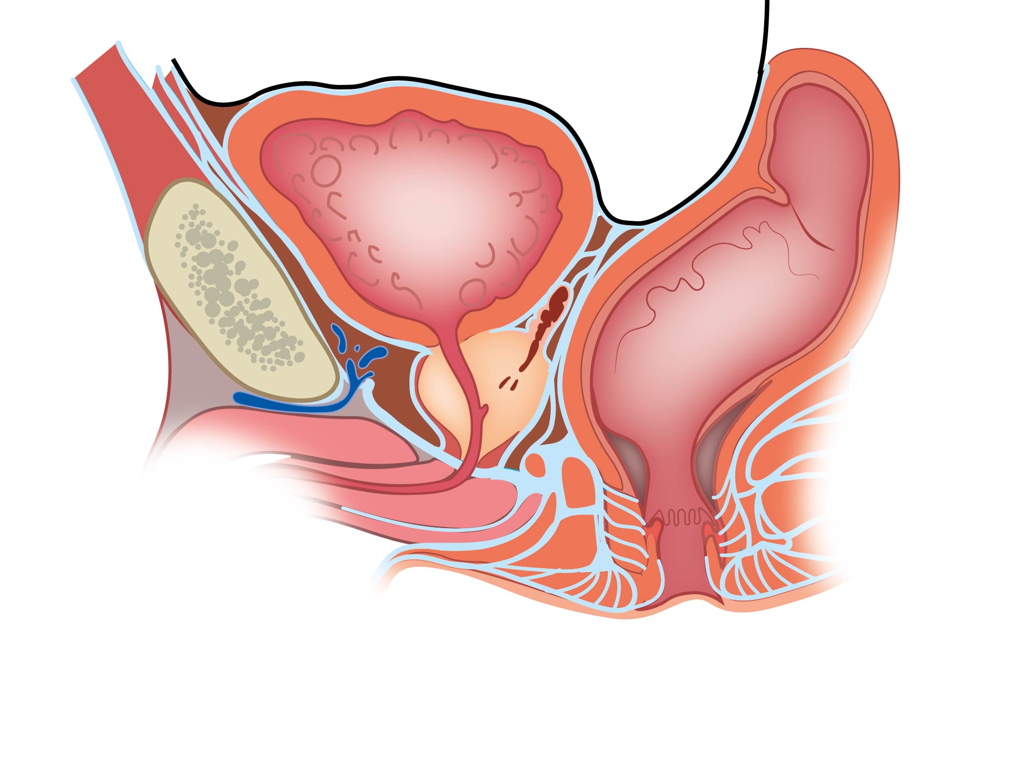
Median section of male pelvis. The black line shows the peritoneal cover, with pouch of Douglas between the posterior bladder and anterior rectum. The turquoise lining is pelvic fascia and the pelvic membrane. The prostate and seminal vesicle are shown inferior to the bladder, superior the perineal membrane. The venous structure retropubic is Santorini's plexus. No labels.
Illustration by Ron Slagter and Marco DeRuiter for course 'Surgical Anatomy of the lesser pelvis' by the 'Urologisch Opleidings Instituut', the Netherlands.
Illustration by Ron Slagter and Marco DeRuiter for course 'Surgical Anatomy of the lesser pelvis' by the 'Urologisch Opleidings Instituut', the Netherlands.
Anatomical structures in item:
Uploaded by: Siem Zethof
Netherlands, Leiden – Leiden University Medical Center, Leiden University
Excavatio vesicouterina
Prostata (Glandula prostatica)
Vesicula seminalis
Membrana perinei
Pelvis
Creator(s)/credit: Ron Slagter NZIMBI, medical illustrator, LUMC; Prof. Marco DeRuiter PhD, anatomist, LUMC
Requirements for usage
You are free to use this item if you follow the requirements of the license:  View license
View license If you use this item you should credit it as follows:
- For usage in print - copy and paste the line below:
- For digital usage (e.g. in PowerPoint, Impress, Word, Writer) - copy and paste the line below (optionally add the license icon):
"Median section of male pelvis – no labels " at AnatomyTOOL.org by Ron Slagter, LUMC and Marco DeRuiter, LUMC, license: Creative Commons Attribution-NonCommercial-ShareAlike
"Median section of male pelvis – no labels " by Ron Slagter, LUMC and Marco DeRuiter, LUMC, license: CC BY-NC-SA


Comments