
nid: 59141
Additional formats:
None available
Description:
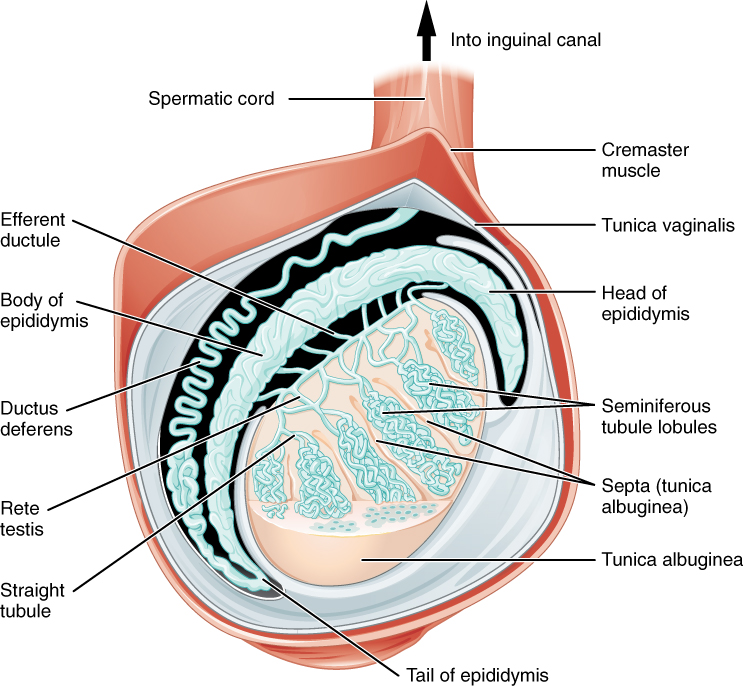
Anatomy of the Testis. This sagittal view shows the seminiferous tubules, the site of sperm production. Formed sperm are transferred to the epididymis, where they mature. They leave the epididymis during an ejaculation via the ductus deferens. English labels. From OpenStax book 'Anatomy and Physiology', fig. 27.4.
Anatomical structures in item:
Uploaded by: Jorn IJkhout
Netherlands, Leiden – Leiden University Medical Center, Leiden University
Colliculus inferior
Funiculus spermaticus
Efferent ductule
Epididymis
Corpus epididymidis
Ductus deferens
Rete testis
Tubuli seminiferi recti
Cauda epididymidis
Tunica albuginea testis
Tubuli seminiferi contorti
Caput epididymidis
Tunica vaginalis testis
Musculus cremaster
Creator(s)/credit: OpenStax
Requirements for usage
You are free to use this item if you follow the requirements of the license:  View license
View license If you use this item you should credit it as follows:
- For usage in print - copy and paste the line below:
- For digital usage (e.g. in PowerPoint, Impress, Word, Writer) - copy and paste the line below (optionally add the license icon):
"OpenStax AnatPhys fig.27.4 - Testis - English labels" at AnatomyTOOL.org by OpenStax, license: Creative Commons Attribution. Source: book 'Anatomy and Physiology', https://openstax.org/details/books/anatomy-and-physiology.
"OpenStax AnatPhys fig.27.4 - Testis - English labels" by OpenStax, license: CC BY. Source: book 'Anatomy and Physiology', https://openstax.org/details/books/anatomy-and-physiology.


Comments